4718
A time efficient, high resolution 3D vessel wall MRI (vwMRI) of the head and neck vessels in a single scan1UCAIR, Department of Radiology and Imaging Sciences, University of Utah, Salt Lake City, UT, United States, 2Department of Neurology, University of Utah, 3Siemens Healthcare, 4Department of Veterans Affairs, VASLCHCS
Synopsis
Vessel wall MRI(vwMRI) increases diagnostic accuracy for stroke etiologies without overt luminal irregularities, such as mild atherosclerosis, subtle dissection, or vasculitis. Prior vwMRI research has focused on the head or neck in isolation. We developed a set of two neck-shape-specific(NSS) coil that fit two different neck sizes and configured them to integrate with the existing commercial head coils. The purpose of this work was to develop a 3D vwMRI protocol, leveraging the NSS coil array, which permits simultaneous imaging of the head and neck vessels in a single scan. By allowing an efficient examination and identifying patients at high risk of future recurrent stroke, this technique will enable detection of cryptogenic stroke sources and optimal personalized management of vascular disease.
PURPOSE
Compared to traditional luminal angiographic techniques, vessel wall MRI provides imaging of both the arterial lumen and, by suppressing MRI blood signal, the vessel wall itself. This increases diagnostic accuracy for stroke etiologies without overt luminal irregularities, such as mild atherosclerosis, subtle dissection, or vasculitis 1-4. Prior vessel wall MRI research has focused on the head or neck in isolation, which is incomplete and inefficient. We developed a set of two high-performance neck-shape-specific (NSS) coil arrays that fit two different neck sizes and configured them to integrate with the existing commercial head coils replacing the lower SNR OEM anterior neck array 5. This approach allowed time-efficient, high-resolution pulse sequences to simultaneously evaluate vascular disease in the head and neck within a single scan. The purpose of this work was to develop a 3D vessel wall MRI (vwMRI) protocol, leveraging the NSS coil array, which permits simultaneous imaging of the head and neck vessels in a single scan.METHOD
The NSS coils are interchangeable to fit different neck sizes. Each array operates simultaneously with the standard high-performance Siemens 3T head coils in our institution. Four different internally developed 3D motion insensitive Stack of Stars (SoS) sequences were used in this study, including SLIPR, 3D IR-SoS, 3D DW-DE SoS and meSoS-DCE 6-8. To utilize the efficiencies in image acquisition using NSS coils we optimized all sequences in our vwMRI protocol to enable complete head and neck coverage in a single acquisition. Pre and post contrast acquisitions were included in the optimized protocol which required only a single injection as opposed to the previous two. Patients with atherosclerotic disease or stroke were imaged using the vwMRI protocol and NSS coils on a 3T MRI scanner (MAGNETOM Prisma, Siemens Healthcare, Erlangen, Germany).RESULT
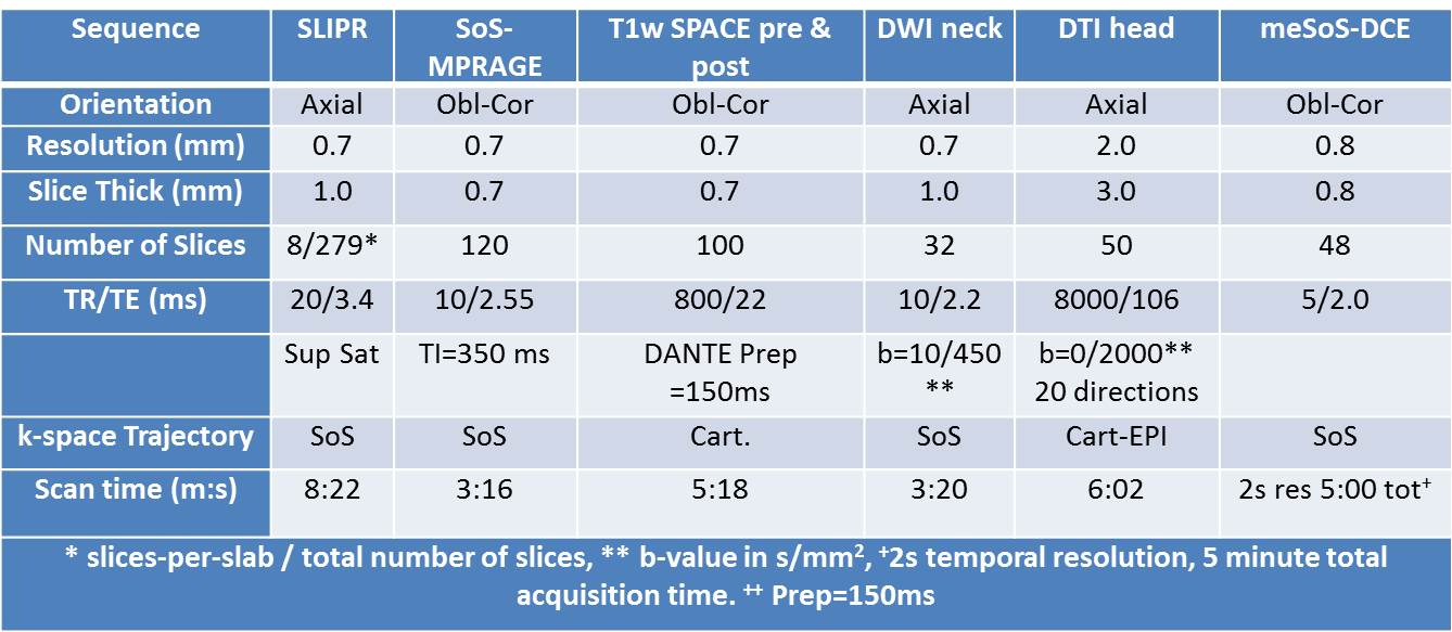
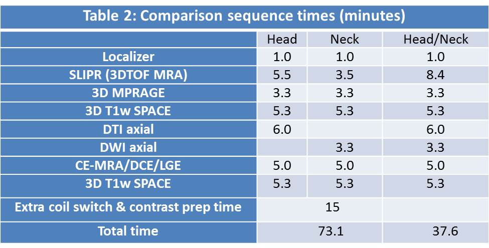
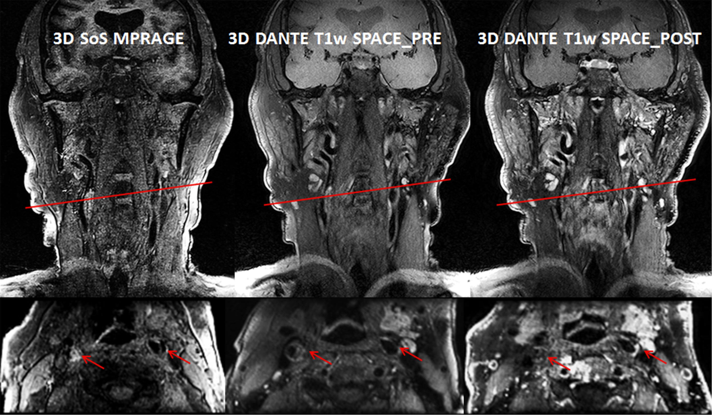
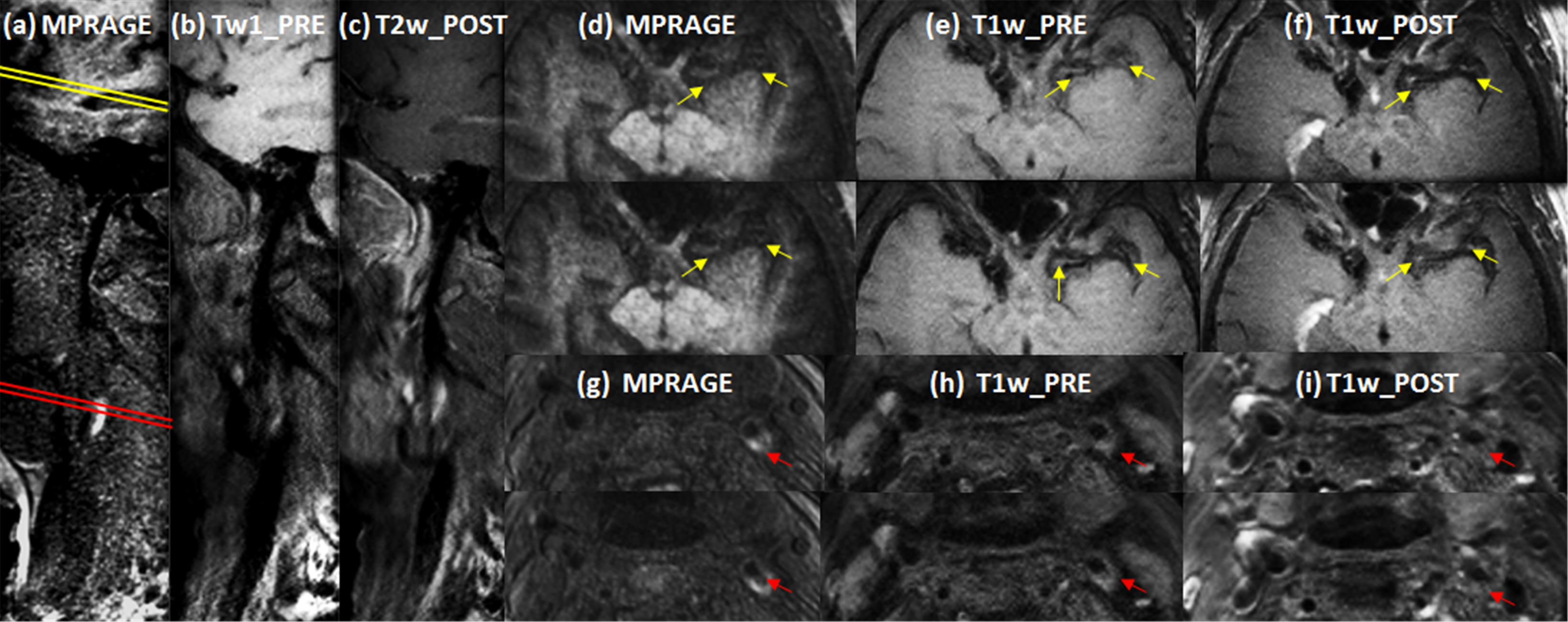
Table 1 summarizes the 3D vwMRI protocol. All sequences, except DWI and DTI, were configured to cover the common carotid artery through the Circle of Willis with the same (0.7 mm) spatial resolution, allowing evaluation of vascular disease in all major vessels feeding the brain. DTI in the brain was used to detect recent stroke. The total exam time including contrast injection was less than 40 minutes.Table 2 displays the comparison sequence times between 3D vwMRI in a single scan and separate head and neck scans. Our protocol reduced the total scan time by 51% compared to separate head and neck scans for the same coverage. Fig 1 shows curved multiplanar reconstructions (MPRs) from SoS-MPRAGE, T1w SPACE pre and post contrast images (top) with zoomed images (bottom) in transverse view of carotid plaque(marked by red arrows) at the red lines drawn on coronal MPRs. Vessel wall and plaque components including calcification and intraplaque hemorrhage(IPH) were well visualized. Fig 2 shows the sagittal MPRs of MPRAGE(a), T1w SPACE pre(b) and post(c) with transverse views at the intracranial (yellow lines) and carotid (redline) from a patient who has both intracranial and carotid plaques. The MPRAGE sequence demonstrates carotid IPH as bright signal(g). Intracranial T1_post images(f) demonstrate contrast leakage into plaque, which indicates inflamed atherosclerosis and a 30% yearly risk of stroke recurrence9.DISCUSSION
Using this technology, it is possible to perform vwMRI examinations of the major head and neck vessels in less than 51% of the time required to image the head and neck separately with a coil switch in between. This is accomplished with the same resolution and improved signal-to-noise. Up to 1/3 of all strokes are considered cryptogenic, meaning that a thorough diagnostic workup failed to find a stroke etiology10. The NSS coils and the availability of efficient vwMRI protocols for the head and neck should greatly improve the ability to detect stroke etiology. By allowing an efficient examination of the arteries feeding the brain, this technology will immediately impact patient. By identifying patients at high risk of future recurrent stroke, simultaneous head and neck vwMRI will enable detection of cryptogenic stroke sources and optimal personalized management of vascular disease.Acknowledgements
Supported by by R01 HL127582, Siemens Medical Solutions, RSNA Research Scholar Grant RSCH1414, and the Clinical Merit Review Grant from the Veterans Administration Health Care System.References
1. de Havenon A, Yuan C, Tirschwell D, et al. Nonstenotic Culprit Plaque: The Utility of High-Resolution Vessel Wall MRI of Intracranial Vessels after Ischemic Stroke. Case Reports in Radiology 2015;2015:e356582.
2. McNally JS, Yoon HC, Kim SE, et al. Carotid MRI Detection of Intraplaque Hemorrhage at 3T and 1.5T. J Neuroimaging 2015;25(3):390-396.
3. Gupta A, Baradaran H, Schweitzer AD, et al. Carotid Plaque MRI and Stroke Risk A Systematic Review and Meta-analysis. Stroke 2013;44(11):3071-3077.
4. Mossa-Basha M, Hwang WD, De Havenon A, et al. Multicontrast high-resolution vessel wall magnetic resonance imaging and its value in differentiating intracranial vasculopathic processes. Stroke; a Journal of Cerebral Circulation 2015;46(6):1567-1573
5. Beck MJ, Parker DL, Bolster BD, et al. Interchangeable Receive-Only Carotid Coils for Simultaneous Imaging with Radio Frequency Head Coils at 3 Tesla. Proceedings of the 24th ISMRM, Singapore, 2016.
6. Kim SE, Roberts JA, Eisenmenger LB, et al. Motion-insensitive carotid intraplaque hemorrhage imaging using 3D inversion recovery preparation stack of stars (IR-prep SOS) technique. J Magn Reson Imaging. 2016 Jul 7. doi: 10.1002/jmri.25365. [Epub ahead of print]
7. Kim SE, McNally JS ,Treiman GS, et al. Motion Insensitive high resolution in vivo DWI of Carotid Artery Wall Imaging using 3D Diffusion Weighted Driven Equilibrium Stack of Stars (3D DW-DE SOS) sequence, Proceedings of the 24th ISMRM, Singapore, 2016.
8. Mendes JK. McNally S, Kim SE, et al. Identifying Carotid Plaque Inflammation Using High and Low Molecular Weight Contrast Agents Proceedings of the 24th ISMRM, Singapore, 2016.
9. Kim JM, Jung KH, Sohn CH, et al. Intracranial plaque enhancement from high resolution vessel wall magnetic resonance imaging predicts stroke recurrence. Int J Stroke. 2016;11:171–179.
10. Li L, Yiin GS, Geraghty OC, Schulz UG, et al. Incidence, outcome, risk factors, and long-term prognosis of cryptogenic transient ischaemic attack and ischaemic stroke: a population-based study. The Lancet Neurology. 2015;14:903–913.
Figures