4716
Accelerated Intracranial Vessel Wall Imaging using Compressed Sensing1Radiology, University of California, San Francisco, San Francisco, CA, United States, 2Radiology, Changhai Hospital, Shanghai, People's Republic of China, 3Siemens Healthcare, CA, United States, 4Siemens Healthcare, Erlangen, Germany
Synopsis
Current 3D black-blood high-resolution MRI (0.4-0.6mm isotropic) of intracranial vessel wall is limited by long scan times (~10 minutes). This study implemented a compressed sensing SPACE (CS-SPACE) sequence to reduce the scan time. The scan and reconstruction parameters were optimized in volunteers and then validated in patients. The optimized CS-SPACE protocol achieved good image quality and reliable vessel area measurements compared with SPACE, with a 37% time reduction. 0.5mm isotropic resolution can be achieved in <7 minutes, and 0.6mm3 is possible in 4 minutes. This fast intracranial vessel wall technique has potential for use in a clinical setting.
Purpose
3D high resolution black blood MRI has been widely used for imaging of vessel wall pathology of intracranial vascular diseases, including atherosclerotic plaques, aneurysms and vasculitis. High isotropic resolution (0.4mm to 0.6mm) has been achieved with good blood suppression and image quality. However, these protocols often need a long scan time of around 10 minutes, which is poorly tolerated by patients 1-2. Acceleration techniques that significantly reduce the scan time while maintaining image quality would be very valuable. This study aims to evaluate and optimize compressed sensing methods for fast intracranial vessel wall imaging.Methods
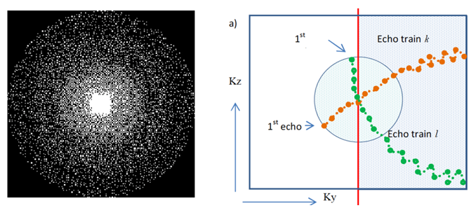
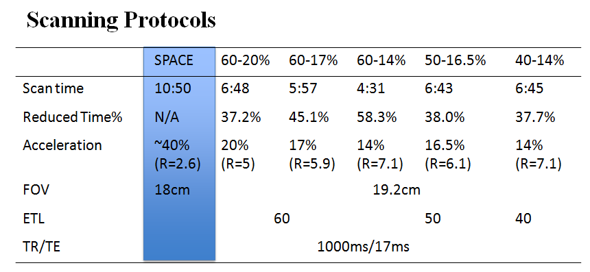
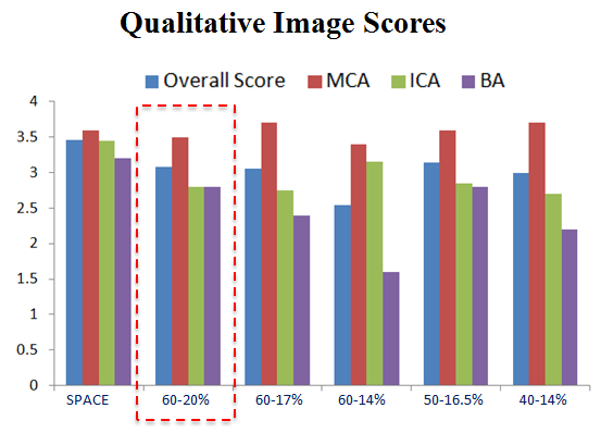
Sequence: A compressed sensing fast-spin-echo sequence (CS-SPACE) was implemented on a Siemens 3T scanner. A Poisson-disk k-space pattern and symmetric radial k-space sampling trajectory were used (Figure 1). Online reconstruction was implemented and took ~2 minutes (20 iterations) for current CS-SPACE protocols. The scan and reconstruction parameters were first optimized in 5 healthy volunteers, and then the optimized protocol was evaluated on 5 patients with cerebral vascular disease (3 intracranial aneurysms, 1 vertebral artery dissection, and 1 vertebral artery occlusion.) Patients were scanned with SPACE and CS-SPACE both pre- and post-Gd contrast injection. The original SPACE has whole brain coverage and 0.5mm isotropic resolution (Table 1). CS-SPACE scanning parameter optimization: Echo train length from 40 to 60, and under-sampling factors from 14% to 20% were tested. Reconstruction parameter optimization: Iteration times from 10 to 40 and regularization factor from 0.0005 to 0.004 were tested. Image analysis: For volunteers, three major intracranial arteries (bilateral middle cerebral and internal carotid arteries, and basilar artery) were included in analysis. Qualitative scores were graded by an experienced radiologist (0-4 scale). Quantitative area and contrast ratio (wall/lumen) were measured. Image scores were evaluated in both sagittal and axial planes. For patients, the qualitative image quality and characterization of contrast enhancement were evaluated.Results
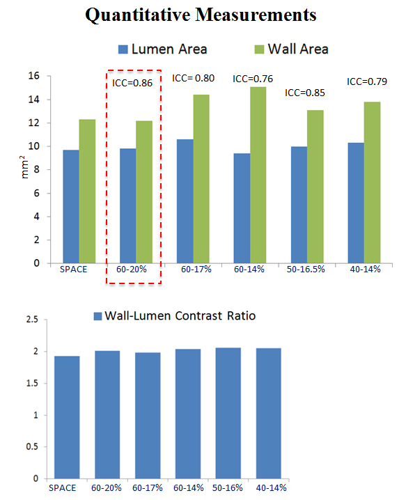
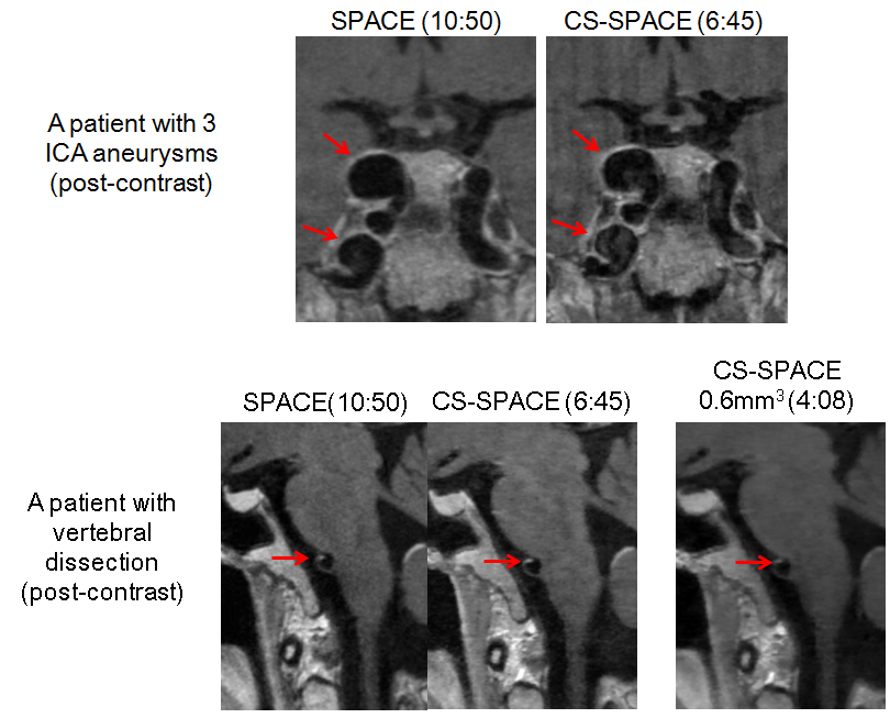
The quantitative comparison is shown in Figure 2 and qualitative image scores are shown in Figure 3. Generally there is good agreement for area measurements between CS-SPACE and SPACE, and there is no difference in wall-lumen contrast ratio. The highest performing scanning protocol has ETL 60 and 20% under-sampling factor. Reconstruction tests showed 20 iterations times and regularization factor of 0.002 provided the best image quality. The optimized protocol achieved comparable image quality to the fully sampled SPACE with a scan time reduction of 37% (6:48 vs. 10:50). Sample patient images are shown in Figure 4. There is no significant difference in the image quality between CS-SPACE and SPACE. SPACE and CS-SPACE had perfect agreement on identifying wall enhancement (3 of 5 patients demonstrated enhancement). We also tested a 0.6mm isotropic protocol which can be done in ~4 minutes with good image quality.Discussion
To our knowledge, this is the first study to apply compressed sensing to 3D black blood intracranial vessel wall imaging. Our optimized CS-SPACE protocol shows good image quality and reliable area quantification compared to SPACE, in a scan time of less than 7 minutes (37% reduction in scan time). 0.6mm isotropic imaging is possible with CS-SPACE in as little as 4 minutes. Fast imaging of intracranial vessel wall using CS-SPACE is a promising tool to evaluate patients with intracranial vascular disease in the clinical setting, and has the potential to improve risk stratification in these patients.Conclusion
Compressed Sensing SPACE can achieve high isotropic resolution whole brain intracranial vessel wall imaging with comparable image quality to SPACE, while significantly reducing the scan time. This technique has the potential to be used in a clinical setting for the evaluation of patients with cerebral vascular diseases.Acknowledgements
This study is supported by National Institute of Health (NIH) grants R01HL114118 and R01NS059944References
1. Fan Z et al, Whole-brain intracranial vessel wall imaging at 3 Tesla using cerebrospinal fluid-attenuated T1-weighted 3D turbo spin echo. MRM 2016
2. Zhu C et al, High resolution imaging of the intracranial vessel wall at 3 and 7 T using 3D fast spin echo MRI. MAGMA 2016
Figures