4711
A preliminary study on the amplitude of low frequency fluctuations in CADASIL1Shanghai Key Laboratory of Magnetic Resonance and Department of Physics, School of Physics and Materials Science, East China Normal University, Shanghai, People's Republic of China, 2Department of Neurology and Jiuyuan Municipal Stroke Center, Shanghai Ninth People's Hospital, Shanghai Jiao Tong University School of Medicine, Shanghai, People's Republic of China
Synopsis
The purpose of this study was to investigate spontaneous low-frequency fluctuations in CADASIL patients during resting-state fMRI scans. Eleven patients (aged, 33-66 years, 6 female) and 11 age- and gender-matched healthy controls were recruited. Amplitude of low-frequency fluctuation (ALFF) was calculated to measure spontaneous brain activity. The results showed that CADASIL patients exhibited significantly decreased ALFF in the bilateral precuneus, and increased ALFF in the midbrain/ pons, the insula/ temporal pole, and the anterior cingulate gyrus/ corpus callosum. Our study first provides empirical evidence for altered spontaneous neuronal activity in CADASIL patients.
Purpose
Cerebral autosomal-dominant arteriopathy with subcortical infarcts and
leukoencephalopathy (CADASIL) is an autosomal dominant vascular disorder. The
disorder manifests clinically as a diffuse leukoencephalopathy with liability
to cognitive impairment, recurrent stroke, and mood disturbance. Recently, functional
magnetic resonance imaging (fMRI) has been used to research the dysfunction in
CADASIL1,2; however, information regarding spontaneous low-frequency
fluctuations in CADASIL was limited. The aim of this study was to investigate amplitude
of low-frequency fluctuation (ALFF) in CADASIL patients under resting-state.Materials and Methods
Eleven subjects diagnosed with CADASIL (aged, 33-66 years, 6 female) and 11 age- and gender-matched healthy controls participated in this study. Functional images were acquired using an EPI sequence with the following parameters: TR/ TE = 2000/ 30 ms, 210 volumes. Structural scans included a high-resolution three-dimensional T1-weighted magnetization-prepared rapid-acquisition gradient-echo pulse sequence (TR/ TE = 2530/ 2.34 ms, 192 slices) and a fluid-attenuated inversion recovery sequence (FLAIR; TR/ TE = 9000/ 93 ms, 30 slices). We calculated ALFF (0.01~0.08 Hz) to measure spontaneous brain activity using Data Processing Assistant for Resting-State fMRI software. Two-sample t test was used to determine between-group differences by SPM12. Moreover, the mask of white matter hyperintensities (WMH) was generated from the skull-stripped FLAIR images using ImageJ software by applying a threshold on signal intensity derived from the signal intensities histogram3. And then the total volume of WMH was normalized (nWMH; normalized WMH volume) to intracranial cavity volume in each patient, which was calculated on the T1-weighted images. The correlation was computed between the nWMH and ALFF in the brain regions showing altered ALFF in patients.Results
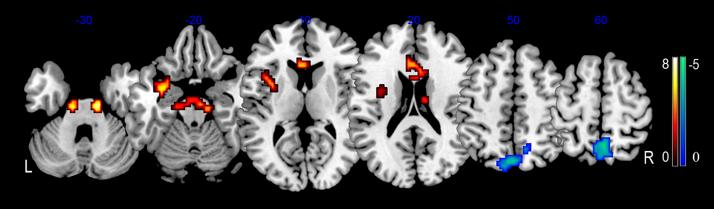
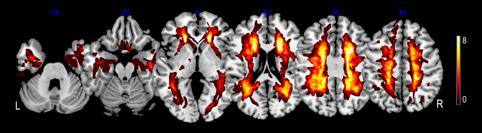
When compared to healthy controls, CADASIL patients exhibited significantly decreased ALFF in the bilateral precuneus, and increased ALFF in the bilateral midbrain/ pons, the left insula/ temporal pole, and the bilateral anterior cingulate gyrus/ corpus callosum, see Figure 1. Furthermore, nWMH = 5.6% ± 2.1% (mean ± SD), and WMH were mainly located in periventricular area, deep whiter matter and temporal pole, see Figure 2. We did not find significant correlations between nWMH and ALFF in patients.Discussion
The decreased ALFF in the bilateral precuneus were observed in patients with CADASIL, which may be associated with cognitive deficit1. It is well known that precuneus was involved in several cognitive functions, for example, episodic memory and visuospatial processing. In addition, a previous study reported2 that patients with CADASIL showed greater radial diffusivity value in the midbrain and pons. Thus, the increased ALFF in the midbrain/ pons could be attributed to the microstructural abnormalities in patients. Given the relatively small sample size, further studies with a larger sample would be necessary to make our conclusion more reliable. Our study first provides empirical evidence for altered spontaneous neuronal activity in CADASIL patients, which may implicate the underlying neurophysiological mechanism in CADASIL.Acknowledgements
This work was supported by grants from the National Natural Science Foundation of China (Nos. 81571658 and 81201082 to X. X. Du)References
1. Cullen B, Moreton F C, et al. Resting state connectivity and cognitive performance in adults with cerebral autosomal-dominant arteriopathy with subcortical infarcts and leukoencephalopathy. J Cereb Blood Flow Metab. 2016; 36(5): 981 - 991.
2. Mascalchi M, Pantoni L, et al. Diffusion Tensor Imaging to Map Brain Microstructural Changes in CADASIL. J Neuroimaging. 2016; doi:10.1111/jon.12374.
3. Viswanathan A. Blood pressure and haemoglobin A1c are associated with microhaemorrhage in CADASIL: a two-centre cohort study. Brain. 2006; 129(Pt 9): 2375 - 2383.
*Correspondence to: Xiaoxia Du. E-mail: xxdu@phy.ecnu.edu.cn.
Figures