4700
Extraction of Dentatorubrothalamic Fiber Tracts in Human Brain Using Probabilistic Fiber Tracking and Unsupervised Clustering Algorithm1Diagnostic Imaging, St.Jude Children's Research Hospital, Memphis, TN, United States
Synopsis
We proposed a novel robust method to extract the dentatorubrothalamic tract (DRTT) in the human brain from DTI images. First, the method was tested on healthy control subjects, then on 30 medulloblastoma patients who had undergone resection of their posterior fossa tumors. Patterns of bilateral, left only, right only and even no DRTT were observed. Validation with multiple manual ROIs along the known pathway confirmed the extracted DRTTs. This suggests that the proposed method may provide an object way to access the DRTTs in medulloblastoma patients post-surgery.
Introduction
Dentatorubrothalamic tract (DRTT) is an important neural fiber tract that connects cerebellum with thalamus and is believed to coordinate movement as well as playing an important role in cognitive function such as planning, verbal fluency, working memory, abstract thinking and behavior [1]. In patients treated for medulloblastoma, the DRTT may be damaged by the tumor itself or by surgical resection. Anatomically, the DRTT originates from the dentate nucleus in the cerebellum and terminates in the contralateral thalamus. Theoretically the DRTT can be tracked if the starting and terminating ROIs are defined. However, the DRTT is a long and complicated multisynaptic tract requiring the tractorgraphy to be tightly constrained using several manual ROIs along the tract [2]. This is both time consuming and subjective. In this study, we proposed a unique method to extract the DRTTs requiring only the dentate nucleus to be manually defined.Method and Materials
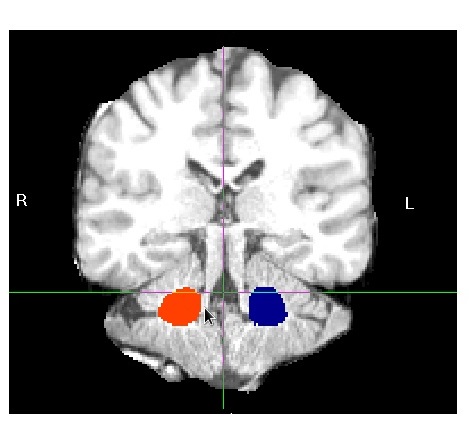
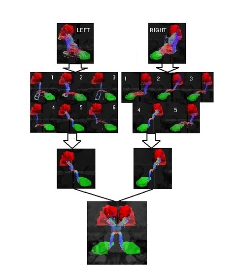
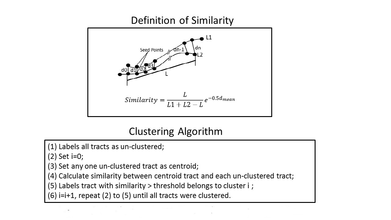
MR scans of 30 childhood medulloblastoma patients (Age at exam 11.2±5.2 years; 8 average-risk [23.4 Gy CRT], 22 high-risk [36.0 Gy CRT]) and 2 healthy adult volunteers (Age 31 and 36) were used in this study. Each MR scan consists of an anatomic 3D T1 weighted image data set and a DTI data set (30 directions, 2 average, b=1000). For each subject, the T1 weighted images were first processed using Freesurfer (http://surfer.nmr.mgh.harvard.edu ) to generate a re-sliced 3D isotropic image (1 x 1 x1 mm) and left and right thalamus masks. The DTI data was pre-processed using the FRMIB Toolbox (http://fsl.fmrib.ox.ac.uk ), and then Bedpost was run to obtain the fiber orientation probability density function on each image voxel for probabilistic fiber tracking. Probtrackx was used to perform fiber tracking. In order to visualize the streamlines of the fibers, the Probtrackx was modified to add a function that can collect each streamline which started from the seed and successfully reached the target, and all the collected streamlines were stored in a format for viewing in Trackvis (http://trackvis.org ). For the DRTT extraction, each side was processed separately taking into account that the tracks start and end in opposite hemispheres. The tracking was performed in the T1 weighted image space and the DTI was non-linearly register to this space. Manual starting ROIs for left and right dentate are shown in Figure 1. The left and right thalamus generated by Freesurfer were used as terminating ROIs. For each seed voxel, 5000 streamlines were generated and each streamline was limited to a maximum length of 150 mm. As shown in Figure 2, the resulting tracks contain both the desired DRTT and other unwanted fibers. To remove the unwanted fibers, a similarity [3] based fiber clustering algorithm (shown in Figure 3) with a clustering threshold of 0.1 was used to group streamlines into several clusters (usually 3 to 10) and the group with shortest mean length was identified as the DRTT. Figure 2 shows the process of clustering of all tracts.Results
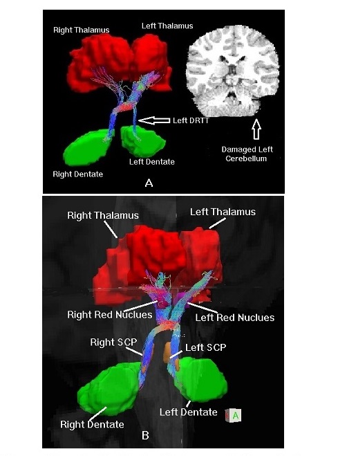
Using the proposed method, the bilateral DRTTs from the volunteer subjects were successfully reconstructed. For the medulloblastoma patients, both left and right DRTTs were successfully reconstructed in 11 subjects; left only DRTTs were reconstructed in 4 subjects; right only DRTTs were reconstructed in 9 subjects; neither DRTT could be reconstructed in 5 subjects. Figure 4 (A) shows the reconstructed bilateral DRTTs of a medulloblastoma patient with damaged left cerebellum. It was noticed that the number of fibers in the left DRTT is less than in the right. By definition, the DRTT is the fiber tract that originates in the dentate, accenting to the same side superior cerebellar peduncle (SCP) and decussating to the contralateral red nucleus (RN) and finally terminating in the contralateral thalamus [4]. To validate our method, left and right SCPs and RNs were manually drawn on the T1 weighted image for 13 subjects (eleven medulloblastoma patients with bilateral DRRTs and two health volunteers) by an independent investigator. As shown in Figure 4 (B), the tracked DRTT fiber bundles were shown to intercept with same side SCP and contralateral RN for all subjects.Discussion and Conclusion
We have described a robust method to extract DRTTs from DTI images requiring only the dentate nucleus to be manually defined. While the DRTTs in medulloblastoma patients are critical fiber tracts that may be damaged by tumor or resection, they cannot be discriminated from adjacent neural structures using only conventional MRI and DTI tractography had previously required multiple time consuming manual ROIs. Therefore, this method may provide an objective feasible way to access damage to the DRTTs in medulloblastoma patients.Acknowledgements
No acknowledgement found.References
[1] Meola A., et al, “The nondecussating pathway of dentatorbrothamic tract in humans: human connectome-based tracktographis study and microdissection validation”, JNS, 124, No.5 1406-1412, 2016
[2] Kwon H. G., et al, “Dentatorunrothalamic tract in human brain: diffusion tensor tractorgraphy study”, Neuroradiology, 53: 787-791. 2011
[3] Ding Z., et al, “Classification Quantification of neuronal fiber pathways using diffusion tensor MRI”, MRM, 49, 716-721, 2003.
[4] Afifi A. K., et al “Functional neuroanatomy: text and atlas”, 2nd edn. Lange Medical Books, McGraw-Hill, New York, 2005.
Figures