4698
Measuring white matter structure in solid tumor survivors: a fixel-based versus voxel-based approach1Department of Pediatric Hematology and Oncology, University Hospitals Leuven, Leuven, Belgium, 2Department of Radiology, University Hospitals Leuven, Leuven, Belgium, 3Centre for the Developing Brain, King’s College London, 4Imaging Biomarker Experts, Icometrix
Synopsis
Neurotoxicity of multi-agent chemotherapy in survivors of solid non-CNS tumors during childhood, has limitedly been investigated. Nowadays, diffusion-weighted imaging (DWI) is implemented in clinical studies to examine potential white matter changes. However, standard voxel-based analyses of diffusion measures such as fractional anisotropy (FA), only provide information about local white matter structure on a voxel-level, but lack specific information about fiber populations within a voxel. Therefore, we compared a fixel-based versus voxel-based group comparison analysis of DWI images in survivors of pediatric solid tumor versus healthy age-matched controls.
PURPOSE
Assessing long-term effects of cancer treatment on white matter microstructure in survivors of solid non-CNS tumors during childhood.BACKGROUND
The neurotoxic adverse effects of chemotherapy treatment have been well documented for leukemia survivors in pediatric oncology1. Recently, also in breast cancer, changes in the white matter microstructure have been reported2. Neurotoxicity of multi-agent chemotherapy in survivors of solid non-CNS tumors during childhood, however, has only limitedly been investigated3. Given that the brain could be most vulnerable to toxicity during childhood, we investigate the white matter microstructure in survivors of solid tumors during childhood. Nowadays, diffusion-weighted imaging (DWI) is implemented in clinical studies to examine potential white matter changes. However, standard voxel-based analyses of diffusion measures such as fractional anisotropy (FA), only provide information about local white matter structure on a voxel-level, but lack specific information about fiber populations within a voxel4. Therefore, fixel-based analysis4 was recently introduced to investigate both within-voxel microscopic fiber density and macroscopic differences in white matter morphology. In this study, we compared the fixel-based versus standard voxel-based approach in survivors of pediatric solid tumors.METHODS
We acquired DWI in survivors of pediatric solid tumors (n=34, age=[16-35], age at diagnosis=[4-18], time since treatment=[2-16] years), and healthy age-matched controls (n=34) on a 3T Philips Achieva MRI scanner with a 32-channel phased-array head coil. The echo-planar, multi-shell diffusion imaging scheme consists of b-values 700, 1000 and 2800 s/mm2, applied along 25, 40 and 75 uniformly distributed gradient directions respectively, complemented by 10 non-weighted (b=0) images. DWI preprocessing including motion and eddy current correction was performed using ExploreDTI5, DWI bias field correction6 and global intensity normalization7 was performed using MRtrix. Whole-brain white matter micro- and macrostructure was analyzed using MRtrix as following:
(1) Fiber
orientation distributions (FODs) were computed using robust constrained
spherical deconvolution8 with a group average response function.
Individual FODs were registered to a population-based FOD-atlas (see Figure 1).
Transformation fields of these registrations were applied for individual
FA-maps as well as fixel maps of apparent fiber density (AFD) and a combined
measure of AFD and fiber cross-section (FDC).
(2) In
order to investigate white matter by using standard FA-maps, a voxel-based
analysis was performed for group comparison of FA, using permutation testing
and threshold-free cluster enhancement.
(3) To
examine potential microscopic and macroscopic white matter damage, AFD and FDC
were compared between both groups using fixel-based non-parametric permutation
testing and connectivity-based fixel enhancement4.
RESULTS
Voxelwise group comparison of FA-maps showed significantly lower FA-values in survivors than in controls for both crossing and non-crossing fibre regions (see regions depicted in blue in Figure 2, p<.05). Similarly, also FDC appears to be smaller in survivors in multiple brain regions (see fixels in Figure 2, p<.05). As could be inferred from Figure 2, the regions of significantly lower FA overlap with regions of significant FDC decrease in survivors. In contrast, with respect to microscopic white matter structure, AFD is reduced in the single-fiber tracts of the cingulum and corpus callosum only (see Figure 3, p<.05).DISCUSSION
Reduced FA and FDC in cancer survivors in multiple brain regions, suggests a pattern of diffuse white matter changes related to chemotherapy during childhood. Similar results have previously reported for survivors of leukemia1 and breast cancer2,9. However, based on these measures, one could not distinguish between white matter changes at microscopic level vs. at macroscopic level. Given that animal studies specifically revealed potential chemotherapy-induced demyelination10, neuroimaging studies that can distinguish between such changes are desirable in measuring the white matter structure in cancer survivors. In this study, fixel-based analysis enabled us to distinguish between potential white matter toxicity at microstructural vs. macrostructural level, while FA-measures correlated with both (AFD & FDC). By combining information from within-voxel microscopic fibre density and macroscopic morphology, these results confirm the lower specificity of FA-measures in comparison to fixel-based analyses. More specifically, with regard to the microscopic fibre density, we found reduced AFD in the cingulum and corpus callosum. These central white matter tracts in the brain, which are highly myelinated, could be particularly vulnerable for demyelination10.CONCLUSION
This study supports the hypothesis of potential demyelination following chemotherapy in single fibre tracts, whereas macroscopic white matter changes were observed globally.Acknowledgements
No acknowledgement found.References
1 Schuitema, I., Deprez, S., Van Hecke, W., Daams, M., Uyttebroeck, A., Sunaert, S., ... & Veerman, A. J. (2013). Accelerated aging, decreased white matter integrity, and associated neuropsychological dysfunction 25 years after pediatric lymphoid malignancies. Journal of Clinical Oncology, 31(27), 3378-3388.
2 Deprez, S., Amant, F., Yigit, R., Porke, K., Verhoeven, J., Stock, J. V. D., ... & Vandenberghe, J. (2011). Chemotherapy-induced structural changes in cerebral white matter and its correlation with impaired cognitive functioning in breast cancer patients. Human brain mapping, 32(3), 480-493.
3 Sleurs, C., Deprez, S., Emsell, L., Lemiere, J., & Uyttebroeck, A. (2016). Chemotherapy-induced neurotoxicity in pediatric solid non-CNS tumor patients: An update on current state of research and recommended future directions. Critical reviews in oncology/hematology.
4 Raffelt, D.; Smith, RE.; Ridgway, GR.; Tournier, JD.; Vaughan, DN.; Rose, S.; Henderson, R.; Connelly, A.Connectivity-based fixel enhancement: Whole-brain statistical analysis of diffusion MRI measures in the presence of crossing fibres. Neuroimage, 2015, 15(117):40-55
5 Leemans, A., Jeurissen, B., Sijbers, J., & Jones, D. K. ExploreDTI: a graphical toolbox for processing, analyzing, and visualizing diffusion MR data. International Society for Magnetic Resonance in Medicine, 2009, 209, 3537
6 Tustison, N.; Avants, B.; Cook, P.; Zheng, Y.; Egan, A.; Yushkevich, P. & Gee, J. N4ITK: Improved N3 Bias Correction. IEEE Transactions on Medical Imaging, 2010, 29, 1310-1320
7 Raffelt D., Tournier J.-D., Rose S., Ridgway G.R., Henderson R., Crozier S., Salvado O., Connelly A. Apparent fibre density: a novel measure for the analysis of diffusion-weighted magnetic resonance images. NeuroImage. 2012;59:3976–3994
8 Tournier, J.-D.; Calamante, F. & Connelly, A. Determination of the appropriate b-value and number of gradient directions for high-angular-resolution diffusion-weighted imaging. NMR Biomedicine, 2013, 26, 1775-1786
9 Deprez, S., Billiet, T., Sunaert, S., & Leemans, A. (2013). Diffusion tensor MRI of chemotherapy-induced cognitive impairment in non-CNS cancer patients: a review. Brain imaging and behavior, 7(4), 409-435.
10 Seigers, R., & Fardell, J. E. (2011). Neurobiological basis of chemotherapy-induced cognitive impairment: a review of rodent research. Neuroscience & Biobehavioral Reviews, 35(3), 729-741.
Figures
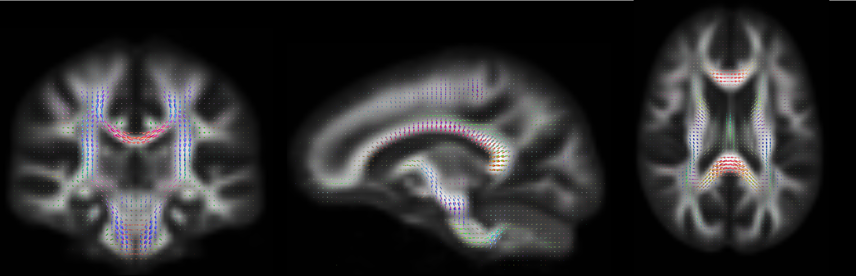
Figure 1. Population-based
atlas for Fiber Orientation Distributions
Fiber Orientation Distributions were computed using robust
constrained spherical deconvolution, with a group average response function
FODs are colored by direction: red = left-right, blue = inferior-superior,
green = anterior-posterior direction

Voxelbased comparison of FA was performed by using permutation testing and threshold-free cluster enhancement,
whereas the fixelbased comparison of FDC was performed by using permutation testing and connectivity-based fixel enhancement
Lower FA in survivors is depicted in blue (p<.05), lower FDC is colored by p-value (p<.05)
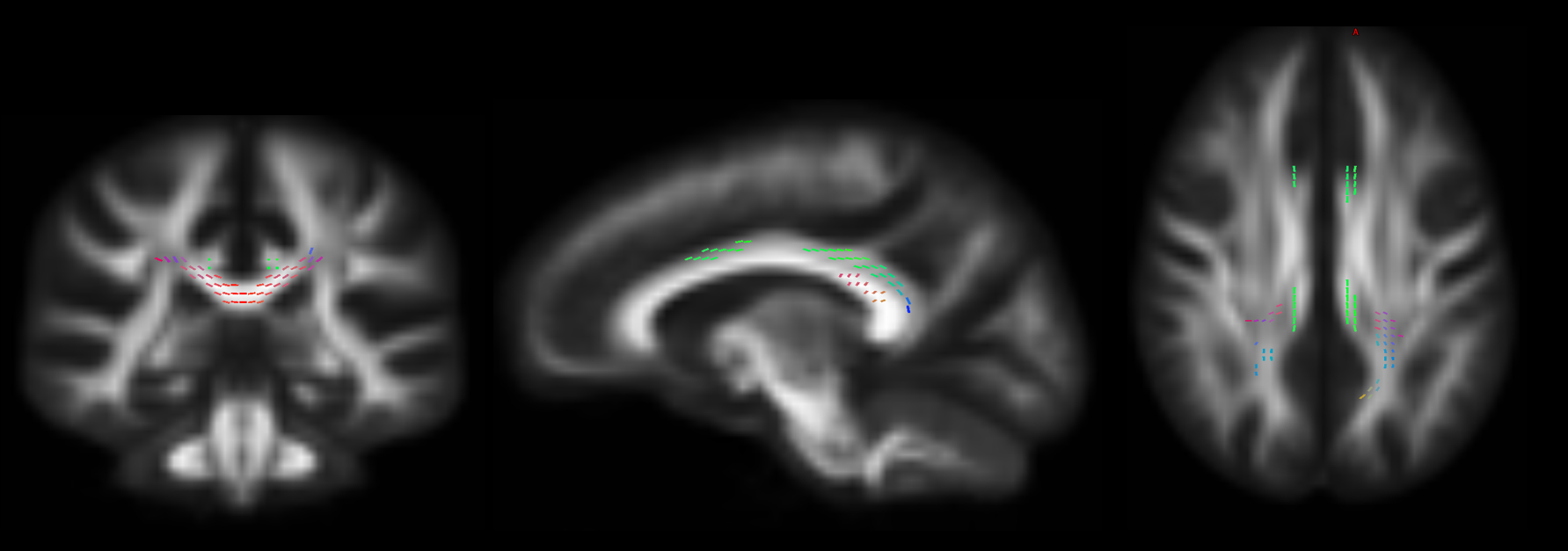
Figure 3. Results
of fixel-based group comparison between survivors and control participants:
comparison of apparent fiber density (AFD)
Lower AFD in survivors is depicted (colored by direction) (p<.05)