4669
3D pituitary dynamic MR imaging using the TWIST and Iterative reconstruction TWIST1Department of Diagnostic Imaging and Nuclear Medicine, Graduate School of Medicine, Kyoto University, Kyoto, Japan, 2Human Brain Research Center, Graduate School of Medicine, Kyoto University, Kyoto, Japan, 3Siemens Healthcare GmbH, Erlangen, Germany
Synopsis
The aim of this study is to perform a comparison between 3D pituitary dynamic study using time-resolved angiography with interleaved stochastic trajectories (TWIST) and TWIST with iterative reconstruction (IT-TWIST). IT-TWIST was retrospectively reconstructed from the raw data of TWIST by using L1 wavelet regularization in space and time. One neuroradiologist put regions of interest in pituitary stalk and anterior lobe and made time intensity curve. The slope of enhancement in the early phase was evaluated. In addition, image quality was visually evaluated. IT-TWIST yielded higher slope of enhancement in the early phase in anterior lobe and better image quality.
Introduction
Materials and Methods
Local institutional review board approved this study.
Patients
Total 8 patients who were suspected seller/paraseller lesions were included in this study. They underwent pituitary dynamic MR study between July 2016 and August 2016. Two of 8 patients were male. The mean age of the included patients was 47.4 years (19–77 years).
MR imaging
Three-dimensional (3D) pituitary dynamic MR imaging using TWIST were acquired on a clinical 3T MR unit (MAGNETOM Skyra, Siemens Healthcare GmbH, Erlangen, Germany) using a 32-channel head coil. Contrast injection was performed with 0.1 mmol/kg patient weight dose of gadolinium based contrast media and 20ml of normal saline was injected at a rate of 4 mL/s 10 s after the scan start via the antecubital vein. TWIST was acquired with interleaved series of central k-space regions A and stochastically undersampled peripheral k-space regions B. Imaging parameters of TWIST were as follows: coronal acquisition; TR/TE, 2.84/1.10 ms; flip angle, 5°; matrix, 203 × 250; field of view, 203 × 250 mm; slice thickness 1mm, isotropic voxel of 1mm, size of the fully sampled central k-space region A, 20%; sampling density of the peripheral k-space region B, 25%; acquisition time, 6.2 s; number of measurements, 15. TWIST images were first directly reconstructed with the product reconstruction after the scan.Afterwards, iterative reconstruction based on prototype was retrospectively performed at the scanner, using a k-t regularization factor of 0.002 and 20 iterations. Reconstruction time was about 8 min employing the graphic processing unit (GPU) of the scanner, and then IT-TWIST images were created.
Imaging analyses
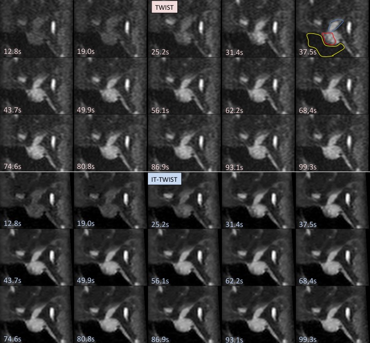
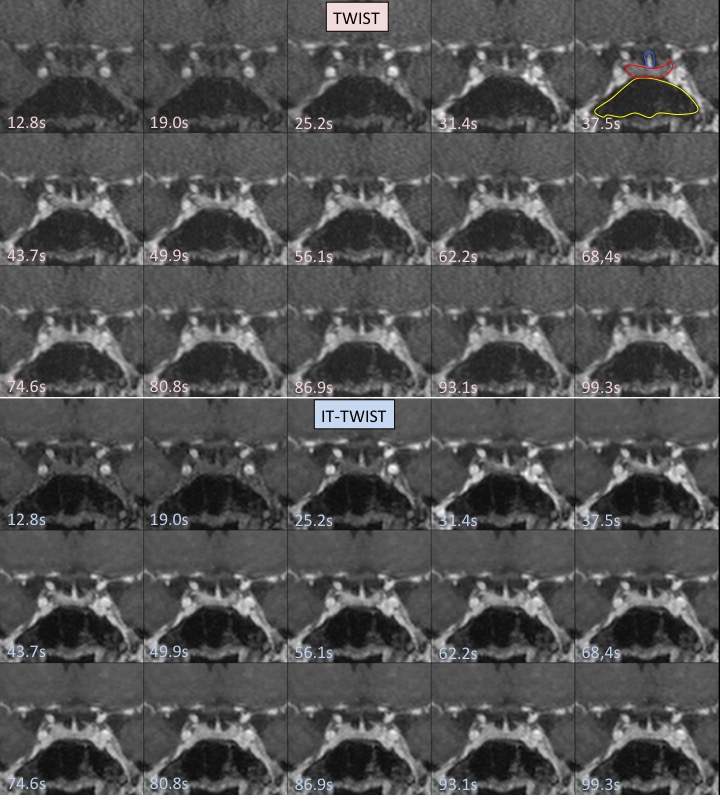
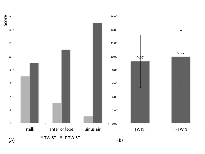
One neuroradiologist visually evaluated the image quality of TWIST and IT-TWIST in terms of delineation of the pituitary stalk and the anterior lobe, and in terms of signal suppression of the air at the sphenoid sinus. Visual evaluation was performed as the following scoring system: comparing TWIST and IT-TWIST, 2 points were given to the better one, and 1 point were given to both images if their quality was equivalent.
Region of interest (ROI) analyses
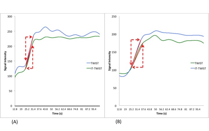
One neuroradiologist placed a circle ROI in the anterior pituitary lobe and the pituitary stalk manually. Time intensity curve (TIC) of TWIST and IT-TWIST images were created for all the patients. TIC was fitted by spline curve method by using Matlab 2014b. The slope of enhancement in the early phase was calculated as follows: Slope = (80% of max value of TIC - 20% of max value of TIC) / Δt. Note that Δt corresponds to the time difference between time at 80% of max value of TIC and time at 20% of max value of TIC).
Results
Discussion
IT-TWIST yielded higher dynamic slope in pituitary dynamic CE study. This retrospective reconstruction method features L1 wavelet regularization in space and time [4] as well as robustness to reduced phase FOVs similar to GRAPPA. TIC of normal pituitary anterior lobe is known to have a higher slope and earlier peak to that of pituitary microadenoma [5]. Higher TIC slope in arterial phase may help differentiate normal pituitary anterior lobe tissue from pituitary adenoma. GPU implementation for faster image reconstruction is achieved with moderate reconstruction time, therefore, clinical application of IT-TWIST retrospective reconstruction become more feasible.
Conclusions
3D pituitary dynamic MR imaging using IT-TWIST showed better visualization of pituitary enhancement compared with that with TWIST.Acknowledgements
This work was supported by Grant-in-Aid for Scientific Research on Innovative Areas “Initiative for High-Dimensional Data-Driven Science through Deepening of Sparse Modeling (No. 4503)” of The Ministry of Education, Culture, Sports, Science and Technology, Japan.References
[1] Wetzl J, Forman C, Wintersperger BJ et al (2016) High-resolution dynamic CE-MRA of the thorax enabled by iterative TWIST reconstruction. Magn Reson Med. 10.1002/mrm.26146
[2] Stalder AF, Schmidt M, Quick HH et al (2015) Highly undersampled contrast-enhanced MRA with iterative reconstruction: Integration in a clinical setting. Magn Reson Med 74:1652-1660
[3] Rapacchi S, Natsuaki Y, Plotnik A et al (2015) Reducing view-sharing using compressed sensing in time-resolved contrast-enhanced magnetic resonance angiography. Magn Reson Med 74:474-481
[4] Liu J, Rapin J, Chang T-c et al (2012) Dynamic cardiac MRI reconstruction with weighted redundant Haar wavelets Proceedings of the 20th Annual Meeting of ISMRM, Melbourne, Australia
[5] M.C. Rossi Espagnet, L. Bangiyev et al. (2015) High-Resolution DCE-MRI of the Pituitary Gland Using Radial k-Space Acquisition with Compressed Sensing ReconstructionAJNR August ; 36(8): 1444–1449.
Figures