4663
Dynamic Contrast-Enhanced MRI Predicts Short-Term Control of Nasopharyngeal Carcinoma within 5 fractions Intensity-Modulated Radiotherapy1Radiology, Fujian Cancer Hospital & Fujian Medical University Cancer Hospital, Fuzhou, People's Republic of China, 2MR Clinical Science, Philips Healthcare, 3Philips Healthcare
Synopsis
Many quantitative DCE-MRI based studies suggested it had utility in early monitoring radiotherapy and chemotherapy sensitivity in anti-tumor treatment. However, there are a few studies investigated whether it could been used for predicting IMRT effect and to what extend of its performance would be. This study enrolled 87 patients who received DCE-MRI one week before NAC and one week after IMRT treatment and suggested there were collaboration the kinetic parameters of quantitative DCE-MRI in early assessing IMRT treatments in NPC.
Purpose Dynamic contrast-enhanced MRI (DCE MRI) had been proved valuable to early predict neoadjuvant chemotherapy (NAC) response in nasopharyngeal carcinoma (NPC) 1. It is suggested that diffusion parameters could be used for predicting Intensity-Modulated Radiotherapy response for NPC 2. However, whether DCE MRI could be used for EARLY assessing IMRT response for locally advanced NPC is remain uncertain. The purpose of this study was To prospectively evaluate DCE-MRI as a tool for assessing short-term control of IMRT in NPC.
Materials and Methods Eeighty-seven newly diagnosed locally advanced NPC patients received two MRI exams using a 3.0 T Philips MRI system (Achieva TX, Best, the Netherlands) one week before treatment and one week after IMRT treatment prospectively. The DCE MRI parameters derived from extended Tofts’ model (including Ktrans [the volume transfer constant of Gd-DTPA], Κep [flux rate constant], νe [the extracellular volume fraction of the imaged tissue], and νp [the blood volume fraction]) were calculated from Philips software developed in IDL 6.3 (ITT Visual Information Solutions, Boulder, CO). Parameters and their corresponding changes △parameter were compared between responders and non-responders after finishing IMRT using independent student T test or Mann-Whitney U test followed by logistic regression and receiver operating characteristic curve (ROC) analyses.
Results Reductions of both Ktrans and Kep values early after one week IMRT were observed in patients who achieved well clinical response after NAC and IMRT treatment. Compared to residual disease (partial response, PR) patients after radical CRT, the pretreatment Ktrans value, percentage change and difference values of Ktrans and Kep parameters between pretreatment and after one week IMRT, and tumor regression ratio after one week IMRT were all significantly larger in complete response (CR) patients (P < 0.05). According to receiver operating characteristic analyses, diagnosis efficacies of single Ktrans, △Ktrans, △Kep, Ktrans(Perc), and Kep(Perc) values ranged from 65.5–82.9%, while combined tumor shrink ratio with above kinetic parameters yielded the highest diagnosis efficacy, sensitivity, and equivalent specificity (Fig 0.832, 84.0%, 64.5%, respectively).
Discussions The DCE MRI derived parameters could reflect physiological features and pathologic changes (hypoxia and angiogenesis) at the micron level 1,3 and corresponding changes after therapy 1. In this pilot study, a combination and consideration of many aspect features of tumor derived from DCE MRI was investigated and proved a higher diagnosis accuracy compare to single kinetic parameter in early predicting IMRT response in locally advanced NPC. Recently, the value of multi-parameters MRI in grading glioma had been validated recently 4.
Conclusions DCE MRI has the potential to predict short-term control of local advanced NPC by means of evaluating changes of tumor vascularization and volume early during treatment. Particularly, the Ktrans value may act as potential markers for predicting the short-term control of NPC toward CRT.
Acknowledgements
No acknowledgement found.References
1. Zheng DC, Chen YB, Liu X, et al. Early response to chemoradiotherapy for nasopharyngeal carcinoma treatment: Value of dynamic contrast-enhanced 3.0 T MRI. J Magn Reson Imaging. 2015; 41(6):1528-40.
2. Hong J, Yao Y, Zhang Y, et al. Value of magnetic resonance diffusion-weighted imaging for the prediction of radiosensitivity in nasopharyngeal carcinoma. Otolaryngol Head Neck Surg. 2013; 149:707-713.
3. Raab P, Hattingen E, Franz K, Zanella FE, Lanfermann H. Cerebral gliomas: diffusional kurtosis imaging analysis of microstructural differences. Radiology. 2010;254(3):876-81.
4. Van Cauter S, De Keyzer F, Sima DM, et al. Integrating diffusion kurtosis imaging, dynamic susceptibility-weighted contrast-enhanced MRI, and short echo time chemical shift imaging for grading gliomas.Neuro Oncol. 2014;16(7):1010-21.
Figures
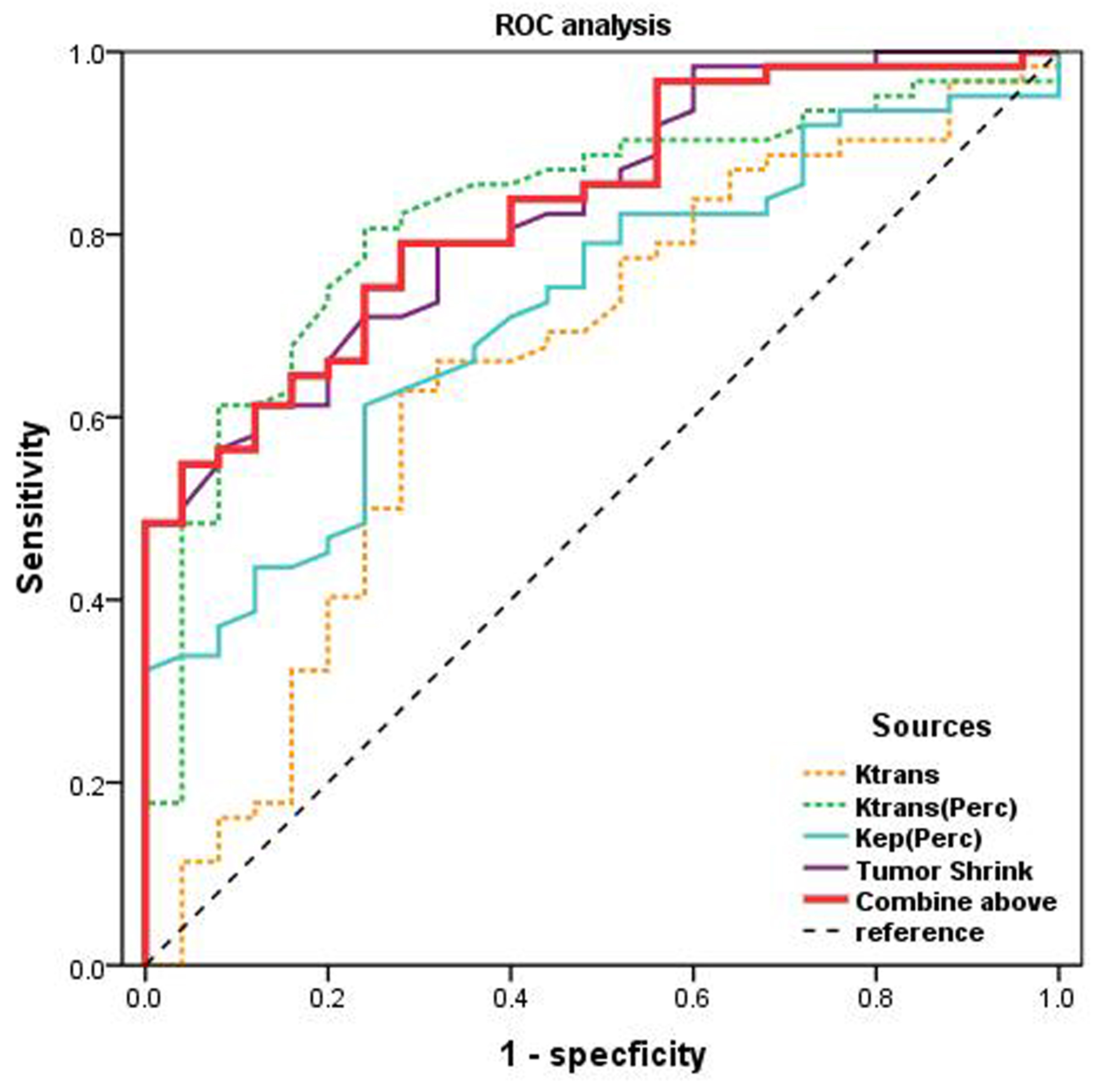
Fig. 1
The diagnostic accuracy of different kinetic parameters (orange: Ktrans, green: Ktrans(Perc), blue: Kep(Perc), purple: Tumor shrink ratio, red: Combine kinetic parameters with tumor shrink ratio, black: reference) in predicting response of CRT of NPC were formed and displayed on figure 1. We concluded that combine kinetic parameters with tumor shrink ratio yielded the highest sensitivity and specificity compared to each alone.