4636
MR Imaging & Spectroscopy in a Non-Human Primate Model of Ebola Makona Aerosol Exposure1NIAID/Integrated Research Facility, National Institutes of Health, Frederick, MD, United States, 2Clinical Research Directorate & Clinical Monitoring Research Program, Leidos Biomedical Research, Inc., Frederick, MD, United States, 3NIAID, Emerging Viral Pathogens Section, National Institutes of Health, Frederick, MD, United States
Synopsis
The purpose of this study was to use MRI and magnetic resonance spectroscopy (MRS) to determine if structural or metabolic alterations occur in the brain of rhesus macaques exposed to Ebola virus via inhalation of aerosolized small particles. Unlike intramuscular inoculation with Ebola virus, small-particle aerosol exposure of macaques did not result in uniform changes in brain volume or vascular alterations 8-9 days after exposure. However, most animals had reductions in N-acetyl aspartate and increases in choline levels, indicating spectroscopy may be useful in identifying early alterations in brain metabolism due to Ebola virus disease.
Purpose
Signs of neurologic disease are difficult to identify in nonhuman primate models of Ebola virus infection. The use of magnetic resonance imaging (MRI) can provide insight to possible structural and metabolic changes occurring during Ebola virus infection. Our previous studies indicated that macaques exposed to 1,000 plaque forming units of Ebola virus (EBOV) by intramuscular injection had significant reductions in whole brain volumes (N=6, -3.3%, p<0.0001), enlarged brain ventricles, as well as alterations in the susceptibility weighted imaging (SWI) indicative of vascular congestion within 7-8 days of exposure[1]. The purpose of the current study was to use MRI and magnetic resonance spectroscopy (MRS) to determine if structural or metabolic alterations occur in the brain of rhesus macaques exposed to Ebola virus via inhalation of aerosolized small particlesMethods
Ten rhesus macaques were exposed to EBOV (Makona variant) via small particle aerosol challenge. Averaged inhaled dose 1150 pfu, with particle size ranging 0.5-3.0 mm. Seven of these animals underwent imaging before exposure and just prior to euthanasia (8-9 days post exposure). Neuroimaging was performed using a clinical 3T Achieva MRI using a pediatric SENSE Head-Spine coil (Philips Healthcare, Andover, MA). Structural brain imaging included: T1-weighted magnetization prepared rapid acquisition with gradient echo (MPRAGE), R2* and T2-weighted images. A 3D MPRAGE sequence was performed that included the following parameters: in-plane resolution of 0.5x0.5x0.5 mm3, TR/TE=9.8/4.7 ms, TI=1100 ms, NSA=2, flip angle of 8o, TFE factor=96, time=4.5 min, and a FOV 96x96x68 mm3. Based on the principle of echo shifting technique [2], a 3D sequence to produce R2* images was created with the following parameters: in-plane resolution of 0.5x0.5x0.5 mm3, TR/TE of 27/35 ms, NSA = 1, flip angle = 5o, time=6 min, and a FOV 96x96x68 mm3. A turbo spin echo (TSE) sequence was used to generate T2-weighted images, which included the following parameters: in-plane resolution of 0.4x0.4 mm2, 2 mm slice thickness, TR/TE=7974/80 ms, NSA=1, fat suppression (SPIR), TSE factor=12, time=4.5 min, and a FOV 96x96x68 mm3. MRS was used to examine changes in brain metabolism in a 1 cm3 region of the gray matter in the fontal cortex. Parameters included: TR/TE=2500/35 ms, the use of a point resolved spectroscopy, 128 averages, chemical shift selective water suppression and the use of 2nd ordered shims. Spectra were fit using LCModel [3]. Paired t-tests were used to determine significant differences between pre and post-exposure metabolite values observed by MRS.Results
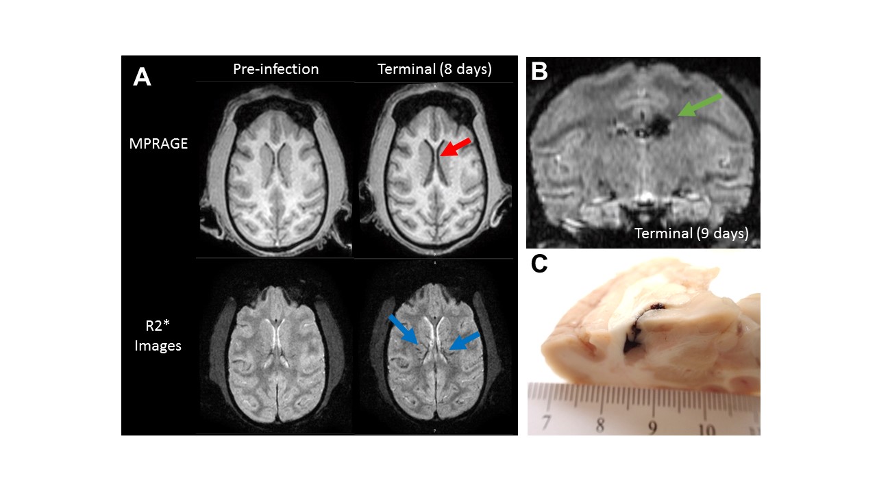
Of the seven animals imaged, only one exhibited reduced brain volume and enlarged ventricles (Figure 1A). Two more animals had indications of vascular changes in the R2* images, one of which was not due to vascular congestion but a focal acute hemorrhage that occurred in the left brain ventricle (Figure 1B). No other significant lesions could be identified by imaging or histologically. While structural MRI can be a useful tool, MRS is far more sensitive, allowing for the detection of neuronal, glial and lipid metabolism that can occur before pathology is evident. Within the frontal cortex (Figure 2), concentrations of N-acetyl aspartate, a marker of neuronal integrity/injury, were found to be reduced in 6 of 7 animals after infection with EBOV/Makona (p<0.01, Figure 3). Choline, a marker of lipid metabolism and suspected marker of glial activation, was found to be elevated in 6 of 7 animals following exposure to EBOV/Mak (p<0.02, Figure 3). Since no significant changes were observed in creatine concentration (p = 0.10), the commonly used metabolite ratios of NAA to creatine (NAA/Cr) and chloline to creatine (Cho/Cr) showed similar changes to that observed in the NAA and choline absolute concentration (Figure 3).Discussion & Conclusion
Unlike IM inoculation with EBOV/Makona, small particle aerosol exposure of macaques to EVOB did not result in uniform changes in brain volume or vascular alterations. However, in many diseases, metabolic changes in tissues are known to precede structural alterations. Reductions in NAA were observed in the frontal cortex (6 of 7 animals), indicating either neuronal injury or relaxation due to increased iron content (vascular congestion) is occurring. Increases in choline levels (observed in 6 of 7 animals) are often attributed to lipid membrane metabolism, glial activation or cellular infiltration in the brain. Further pathologic assessment is required to determine the relationship between alterations in brain metabolism and Ebola virus related disease. In regards to alterations in the brain, these preliminary results indicate that differences exist between intramuscular and aerosol exposure models. Moreover, MRS may be a useful tool in examining early changes in brain metabolism due to Ebola virus disease.Acknowledgements
This work was supported by NIAID Division of Intramural Research and NIAID DCR and was performed under Battelle Memorial Institute contract (No. HHSN272200700016I) with NIAID. Additional support was provided by the NCI Contract No. HHSN261200800001E.References
[1] Lentz MR, et al. “Neuroimaging of Acute Ebola Virus Disease in a Non-Human Primate Model”. International Society of Magnetic Resonance in Medicine, Singapore, 2016.
[2] Moonen CTW, et al. Magn Reson Med. 1992; 26 (1): 184-189
[3] http://s-provencher.com/pages/lcmodel.shtml
Figures