4627
Electric properties tomography in a rodent model of ischemic stroke: Results of a combined ex-vivo and in-vivo pilot study.1Department of Radiology and Neuroradiology, UKSH, Kiel, Germany, 2Department of Experimental and Clinical Pharmacology, UKSH, Kiel, Germany, 3Research Laboratories, Philips GmbH Innovative Technologies, Hamburg, Germany
Synopsis
Electric properties tomography (EPT) is a new contrast in MRI which delivers information on tissue electrical conductivity. Up to now, it has been mostly used for tumor mapping. Ischemic cerebral stroke is another promising application. Seven male Wistar rats were used in this study. Five culled animals from another stroke study, three of which were subjected to MCAO and two live animals which were also subjected to MCAO were examined. Healthy cortical grey matter, white matter and cerebrospinal fluid could be well differentiated Conductivity was altered within the infarct. EPT is feasible in a rodent model of stroke.
Introduction
Electric properties tomography (EPT) is a new contrast in MRI which delivers information on tissue electrical conductivity1. In the clinical realm it has been mostly used for tumor mapping2. Ischemic cerebral stroke with rapidly changing pathophysiology and different compartments (i.e. core, penumbra, oligemia) is another promising but yet almost3 neglected application. It might deliver additional information on tissue viability and possible response to therapy. So far no experiments on experimental stroke have been conducted. The aim of this study was to optimize the sequence and demonstrate the feasibility of EPT in a rodent model of stroke. Further, we aimed to compare electric conductivity in ischemic and non-ischemic cerebral tissue.Material and Methods
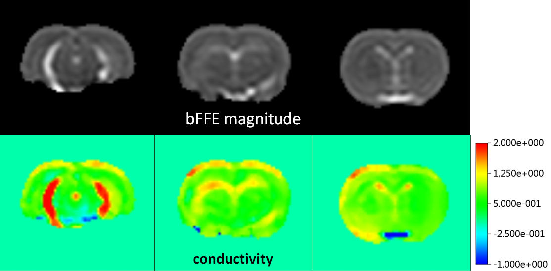
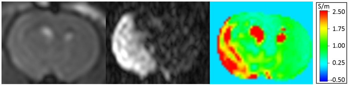
Seven male Wistar rats were used in this study. Five culled animals from another stroke study, three of which were subjected to permanent filament middle cerebral artery occlusion (MCAO) and two live animals which were also subjected to transient (n=1) and permanent (n=1) MCAO were examined. All animals were scanned in a 3T system (Philips Achieva/Best, The Netherlands) using a dedicated solenoid animal coil (Philips/Hamburg, Germany). In addition to DWI, EPT was performed using a Steady-State Free-Precession (SSFP) sequence (TR/TE = 4.5/2.3 ms, measured voxel size = 0.6×0.6×1.2 mm³, flip angle = 38°, NEX = 4). From the transceive phase ϕ of these SSFP scans, conductivity σ was estimated by the equation σ = Δϕ/(2μ0ω) with Δ the Laplacian operator, μ0 the magnetic permeability, and ω the Larmor frequency. Subsequently, a median filter was applied, which was locally restricted to voxels with comparable signal magnitude.Results
All animals subjected to MCAo exhibited an infarct as demonstrated on DWI. Healthy cortical grey matter and white matter showed different conductivity (0.83±0.14 vs. 0.63±0.06 S/m) and could be well differentiated from cerebrospinal fluid (Fig. 1). Conductivity within the infarcted region was 60-70% of the conductivity of healthy grey matter in culled animals (Fig. 2) and in the animal with permanent occlusion but increased in the animal with transient occlusion.Conclusion
EPT is feasible in a rodent model of stroke. Infarcted tissue exhibited altered conductivity. Further in-vivo experiments with examination of the influence of reperfusion status and temporal evolution of the infarcted areas are planned. Depiction of the ischemic penumbra and possibly subcategorisation of the DWI lesion also seem to be a fruitful target for further studies.Acknowledgements
No acknowledgement found.References
1: Katscher U. et al.: Comput Math Methods Med. 2013;2013:546562.
2: Huhndorf M. et al.: In Proceedings of the 21th Scientific Meeting of the International Society of Magnetic Resonance in Medicine 2013:3626.
3: van Lier A.L. et al.: In Proceedings of the 20th Scientific Meeting of the International Society of Magnetic Resonance in Medicine 2012:3484.
Figures