4623
Magnetic particle imaging - The future of acute stroke imaging and treatment?1Neurology, University Medical Center Hamburg-Eppendorf, Hamburg, Germany, 2Institute of Biomedical Imaging, University Medical Center Hamburg-Eppendorf, Hamburg, Germany, 3Neuroradiology, University Medical Center Hamburg-Eppendorf, Hamburg, Germany, 4Materials Science and Engineering Department, University of Washington, Seattle, United States
Synopsis
Magnetic particle imaging (MPI) is a new tomographic imaging modality with superior temporal and spatial resolution compared to other imaging techniques, allowing 3D real-time assessment of vasculature and perfusion without X-rays or nephrotoxic contrast agents. For the first time we show that MPI can used for the diagnosis of acute pathologies like ischemic stroke by showing the first MPI stroke images in a murine stroke model. Additionally, we give an outlook how MPI may revolutionize not only stroke imaging but also stroke treatment, as the magnetic fields of the MPI can be used for catheter guidance and targeted drug delivery.
Introduction
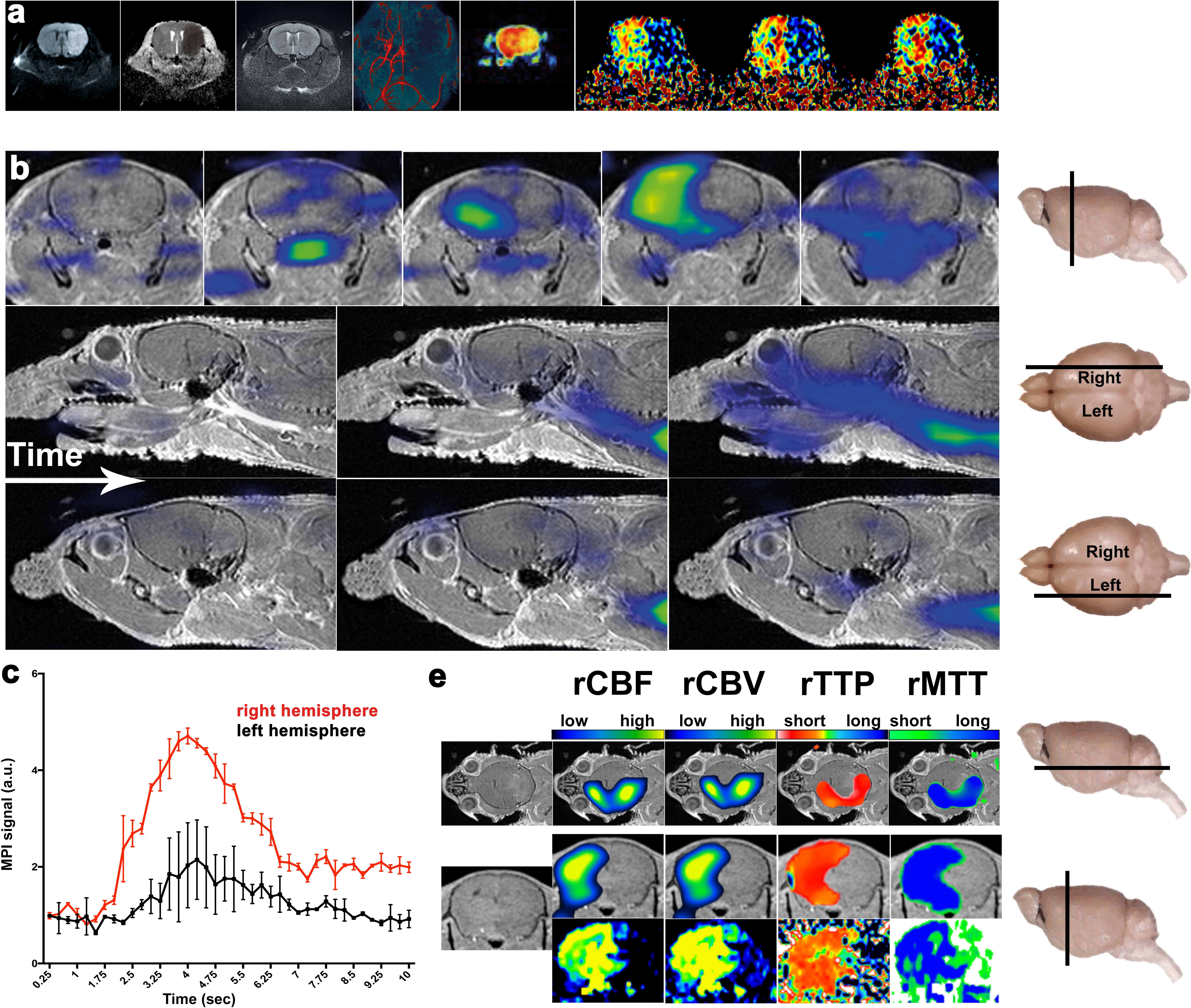
"Time is brain" - with two million neurons dying every minute after stroke onset [1] fast and accurate diagnosis of ischemic stroke is fundamental for successful treatment of stroke patients. Ideally, stroke imaging should be able to distinguish between hemorrhage and ischemic stroke, but also provide further information on the tissue at risk, the so-called penumbra, and stenosis of extra- or intracranial vessels within a short period. Magnetic particle imaging (MPI) is a new imaging modality acquiring 3D datasets combining high spatial resolution with short image acquisition times [2, 3] allowing to perform a broad range of functional cerebrovascular measurements such as cerebral perfusion to detect ischemic stroke in real-time. So far, no MPI- scanners for in vivo experiments have been available. With the first pre-clinical MPI- scanner we address the question, whether MPI is a feasible imaging method for detection of acute pathologies like ischemic stroke. The purpose of this work was to visualize vasculature, vessel occlusion and cerebral perfusion in a murine model of ischemic stroke with MPI.Material and Methods
The left common carotid artery (CCA) was ligated in a 12 week-old C57BL/6 mouse mimicking acute occlusion of this vessel. To assess anatomical information about the murine head and neck, a 7 T small animal MRI was used (Bruker Clinscan). Head position was marked with Resovist (Bayer Schering Pharma AG) filled fiducials for geometry planning and image registration of the MRI and MPI data during post-processing. Magnetic particle imaging was performed using a pre-clinical MPI scanner (Bruker/Philips). A 100 µL bolus with 46 mmol(Fe)/L (LS8, LodeSpin Labs) was injected via the mouse tail vein. MPI scans (3D data with 21.5 ms temporal resolution, a gradient strength of 1.5 Tm-1µ0-1 in two directions, and a drive-field amplitude of 14 mTµ0-1) were acquired dynamically while administering the contrast-agent bolus.Discussion and conclusion
For the first time we show the
capabilities of magnetic particle imaging for the diagnosis of relevant
diseases. In the first preclinical MPI scanner we are able to detect small
perfusion deficits in a murine stroke model with similar precision compared to
MRI. Additionally, several important parameters, as vascular anatomy, stenosis,
and cerebral perfusion can be acquired with a single bolus of contrast agent,
resulting in faster imaging times. Besides imaging, MPI has the potential to
improve acute stroke treatment by using the magnetic fields of the MPI scanner
to manipulate objects. We show first results how MPI can be used for catheter
interventions and exact drug delivery of stroke therapeutics. MPI has the
potential to revolutionize acute stroke imaging and treatment.
Acknowledgements
No acknowledgement found.References
[1] Saver, J.L., Time is brain--quantified. Stroke, 2006. 7(1):p. 263-6.
[2] Buzug, T.K.A.T.M., Magnetic Particle Imaging: An introduction to Imaging Principles and Scanner Instrumentation. 2012: Springer.
[3] Gleich, B. and J. Weizenecker, Tomographic imaging using the nonlinear response of magnetic particles. Nature, 2005. 435(7046): p. 1214-7.
Figures