4609
Correlation of cerebrovascular reserve assessed by acetazolamide-stress SPECT with collaterals on arterial spin-labeling MRI in patients with carotid occlusive disease1Department of Radiology, Seoul Veterans Hospital, Seoul, Korea, Republic of
Synopsis
We evaluated the correlation between cerebrovascular reserve (CVR) on acetazolamide-stress single photon emission computed tomography (SPECT) brain scans and collaterals on arterial spin-labeling (ASL) magnetic resonance imaging (MRI) in internal carotid artery (ICA) stenosis.With acetazolamide stress SPECT, the 21/74 (28%) patients showed evidence of decreased CVR. In 7/53 (13%) of the normal CVR group and 10/21 (48%) of the reduced CVR from the SPECT results, ASL showed ATA in ipsilateral to the stenosis. Significant relationship was observed between reduced CVR group and ATA showing group in ICA stenosis patients on ASL brain perfusion (p=0.004).
Purpose
We evaluated the correlation between cerebrovascular reserve (CVR) on acetazolamide-stress single photon emission computed tomography (SPECT) brain scans and collaterals on arterial spin-labeling (ASL) magnetic resonance imaging (MRI) in internal carotid artery (ICA) stenosis.Methods
74 patients with ICA stenosis (>70%) underwent pulsed ASL brain perfusion scan and SPECT brain perfusion scan in the resting and after acetazolamide challenge. We observed the presence of intracranial collaterals, which are manifested by arterial transit artifact (ATA), on ASL. CVR based on rest-SPECT and acetazolamide-stress SPECT was calculated. With acetazolamide stress SPECT, the 74 patients were grouped as either showing or not showing evidence of decreased CVR. We assessed the relationship between reduced CVR and intracranial collaterals shown as ATA on ASL.Results
In 17/74 (23%) of the ICA stenosis patients, ASL showed ATA in ipsilateral to the stenosis. With acetazolamide stress SPECT, the 21/74 (28%) patients showed evidence of decreased CVR. In 7/53 (13%) of the normal CVR group and 10/21 (48%) of the reduced CVR from the SPECT results, ASL showed ATA in ipsilateral to the stenosis. Significant relationship was observed between reduced CVR group and ATA showing group in ICA stenosis patients on ASL brain perfusion (p=0.004).Discussion
In response to a decrease in local perfusion pressure, there is recruitment of flow from both circle of Willis and leptomeningeal anastomoses, which compensate for the lack of anterograde flow. The eventual failure of collateral pathways is thought to lead to infarct growth and is ultimately responsible for the decreasing efficacy of stroke therapy with time. On ASL, late-arriving flow appears as serpiginous high ASL signal within cortical vessels, which has been termed ATA. ATA with ASL imaging is a region compatible with collateral flow, showing multiple serpiginous high signal intensity structures. ATA was seen frequently in a small group of acute ischemic stroke patients and was associated with tissue survival and improved clinical outcome. Also, patients with chronic hypoperfusion with ATA had good CVR in response to acetazolamide. In our study, statistically significant positive correlation was noted between normal CVR group and ICA stenosis patients with ATA. Patients who had decreased CVR on acetazolamide-challenged SPECT brain studies showed increased dependence on leptomeningeal collaterals.Conclusion
Our results suggest that ICA stenosis patients who had reduced CVR on brain SPECT showed increased dependence on intracranial collaterals shown as ATA on ASL brain scans.Acknowledgements
No acknowledgement found.References
1. Liebeskind DS. Collateral circulation. Stroke. 2003; 34:2279–2284
2. Sheth SA, Liebeskind DS. "Imaging Evaluation of Collaterals in the Brain: Physiology and Clinical Translation". Curr Radiol Rep. 2014 Jan;2(1):29
3. Detre JA, Leigh JS, Williams DS, Koretsky AP. Perfusion imaging. Magn Reson Med. 1992; 23:37-45
4. Zaharchuk G, Do HM, Marks MP, Rosenberg J, Moseley ME, Steinberg GK. Arterial spin-labeling MRI can identify the presence and intensity of collateral perfusion in patients with moyamoya disease. Stroke. 2011 Sep;42(9):2485-2491
5. Smith HA, Thompson-Dobkin J, Yonas H, Flint E. Correlation of xenon-enhanced computed tomography-defined cerebral blood flow reactivity and collateral flow patterns. Stroke 1994; 25:1784–1787 6. Dreyden CP, Grubb RL, Powers WJ. Cerebral hemodynamic impairment: methods of measurement and association with stroke risk. Neurology 1999;53:251–259
7. Derdeyn CP, Shaibani A, Moran CJ, et al. Lack of correlation between pattern of collateralization and misery perfusion in patients with carotid occlusion. Stroke 1999;30:1025–1032
8. Bang OY, Saver JL, Buck BH, Alger JR, Starkman S, Ovbiagele B, et al. Impact of collateral flow on tissue fate in acute ischaemic stroke. J Neurol Neurosurg Psychiatry. 2008; 79:625–629
9. Kidwell CS, Saver JL, Mattiello J, Starkman S, Viñuela F, Duckwiler G, et al. Diffusion-perfusion MRI characterization of post-recanalization hyperperfusion in humans. Neurology. 2001; 57:2015–2021
10. Ye FQ, Berman KF, Ellmore T, Esposito G, van Horn JD, Yang Y, et al. H(2)(15)O PET validation of steady-state arterial spin tagging cerebral blood flow measurements in humans. Magn Reson Med. 2000; 44:450–456
11. Detre JA, Samuels OB, Alsop DC, Gonzalez-At JB, Kasner SE, Raps EC. Noninvasive magnetic resonance imaging evaluation of cerebral blood flow with acetazolamide challenge in patients with cerebrovascular stenosis. J Magn Reson Imaging. 1999; 10:870–875
12. Chalela JA, Alsop DC, Gonzalez-Atavales JB, Maldjian JA, Kasner SE, Detre JA. Magnetic resonance perfusion imaging in acute ischemic stroke using continuous arterial spin labeling. Stroke. 2000; 31:680–687
Figures
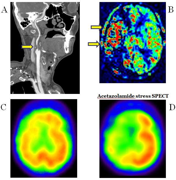
Figure 1. 72-year-old male, severe stenosis of the right proximal cervical ICA.
Multiple serpiginous high signal intensity (red) structures were seen in the right frontotemporal area on pulsed ASL with right proximal cervical ICA stenosis patient, and on acetazolamide stress SPECT image, there was reduced CVR change in the right frontotemporal area.
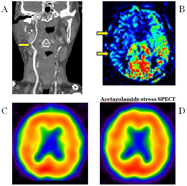
Figure 2. 79-year-old male, severe stenosis of the right proximal cervical ICA.
No ATA in the right frontotemporal area were seen on pulsed ASL with right proximal cervical ICA stenosis patient, and on acetazolamide stress SPECT image, there was no CVR change in the right frontotemporal area.