4603
Disrupted Brain Connectivity Patterns in patients with Attention-deficit/hyperactivity disorder1Department of Radiology, Huaxi MR Research Center, Chengdu, People's Republic of China, 2Department of Psychiatry, The First Affiliated Hospital of Wenzhou Medical University, People's Republic of China
Synopsis
In the current study, we used a graph theory-based network measurements named degree centrality (DC) to identify main cortical hubs in the brain network architecture at voxel level affected in ADHD. Then, functional connectivity maps were generated with seeded at altered DC to detect the brain changes at large-scale level. Finally, we found the disconnection within cortico-thalamus and cortico-striatal loops in ADHD patients, which may associated with inattention and cognitive function deficits.
Introduction
Attention-deficit/hyperactivity disorder (ADHD) is the most commonly diagnosed neurodevelopmental disorder in childhood, characterized as age-inappropriate levels of inattention, impulsivity and hyperactivity. Although brain structural deficit and functional alteration in many cortical regions had been found in patients with ADHD, they do not directly yield information on important functional network topological changes. Here, we aim to identify aberrant brain network architecture in ADHD using a graph theory-based network measurements tool named degree centrality (DC) at the voxel level.Methods
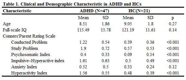
Forty-seven ADHD boys and 21 matched healthy controls were recruited, aged between 6-13 years (Table 1). Behavioral problem was measured using the hyperactivity index from the Chinese version of revised Conners’ Parent Rating Scale (CPRS). CPRS is a battery of questions to evaluate problematic behavior across areas such as sleep, temper and peer relationships, which are aggregated into six factors: Conduct Problem, Study Problem, Psychosomatic, Impulsive–Hyperactive, Anxiety and Hyperactivity Index.
All MRI scans were performed on a GE signal HDx 3T MR scanner with an eight-channel phased-array head coil. Whole-brain resting state fMRI data depicting blood oxygen level-dependent (BOLD) contract were obtained using a gradient-echo echo-planar imaging sequence with the following parameters: 31 axial slices, slice thickness = 4 mm, slice gap = 0.2 mm, repetition time (TR) =2000ms, echo time (TE) =30ms, flip angle = 90°, matrix size = 64×64; field of view (FOV) = 192×192 mm2. Subjects were instructed to relax with their eyes closed without falling asleep during MR examination.
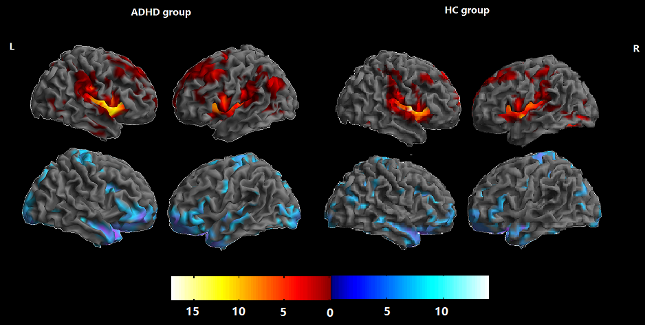
DPARSFA software was used to calculate the parametric maps of degree centrality, a binary matrix was then obtained by thresholding each correlation at p<0.001 (r=0.2). The DC maps of all subjects in each group were analyzed by using a 1-sample t test to identify the spatial distribution of the DC values in each group. The statistical threshold was set at P<.05, with a FDR correction at cluster level. Independent 2-sample t tests were also performed on the basis of the individual DC maps to examine the differences in DC between groups. Then, the identified brain regions with altered DC were chosen as the seeds to examine their functional connectivity (FC) changes.
Results
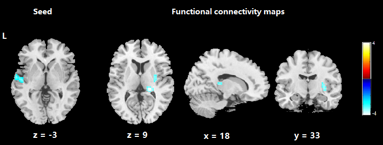
The spatial distribution of the binarize DC maps are mainly located in the insula, basal ganglia, cingulate cortex, temporal gyrus, precuneus and prefrontal cortex in both ADHD and HCs (Figure 1). After two-sample t-test, relative to controls, ADHD patient showed decreased DC in the left superior temporal gyrus (STG) (voxel level p<0.005, Alphsim corrected for cluster level p<0.05). The FC patterns seeded at left STG showed decreased connections with right thalamus and right putamen (voxel level p<0.001, family-wise error correction at cluster level, p<0.01).Discussion
In the current study, we examined whole-brain intrinsic functional architecture in ADHD boys by using a voxel-wise network centrality indices. Compared with HCs, our findings show that the STG is the main cortical hub in the brain network architecture affected in ADHD. Seed-based analytic approaches revealed the brain circuits anchored in thalamus and putamen were also affected, indicating disconnection within cortico-thalamus and cortico-striatal loops.1 These results are consistent with previous task MRI studies demonstrating decreased activation and functional connectivity of the cortico-thalamus and cortico-striatal loops during sustained attention, inhibitory control, and cognitive switching in ADHD relative to HCs.2Conclusion
We found disconnection within cortico-thalamus and cortico-striatal loops in ADHD patients, which may associated with inattention and cognitive function deficits. Future studies on brain functional alterations in ADHD at large-scale level should be conducted to confirm this change.Acknowledgements
This study was supported by the National Natural Science Foundation (Grant No. 81671669 and 81171488)References
1. Leanne T, Vinod M, Allan L. R, Parietal Attentional System Aberrations During Target Detection in Adolescents With Attention Deficit Hyperactivity Disorder: Event-Related fMRI Evidence. Am J Psychiatry 2006; 163:1033–1043
2. Suzanne C, Kurt S, Olga B, et al. Thalamo-Cortical Activation and Connectivity During Response Preparation in Adults With Persistent and Remitted ADHD. Am J Psychiatry 2013; 170:1011–1019
Figures