4600
Traffic-Related Air Pollution Associated with Activation in a Functional MRI Verb Generation Task of a Longitudinally Studied, Pediatric Cohort1Radiology/Imaging Research Center, Cincinnati Children's Hospital Medical Center, Cincinnati, OH, United States, 2Cincinnati Children's Hospital Medical Center, Cincinnati, OH, United States, 3University of Cincinnati College of Medicine, Cincinnati, OH, United States
Synopsis
Traffic-related air pollution (TRAP) is strongly associated with adverse cardiopulmonary health effects. Evidence suggests the developing brain may also be a target organ for particulate matter due to translocation either from the respiratory system or through the olfactory nerve. Using a pediatric cohort, we tested the hypothesis that exposure to TRAP during critical windows of brain development is significantly associated with changes in brain functioning during a language task. Children with high exposure levels were associated with reduced activation within the frontal lobe compared with children at low exposure.
Purpose
Traffic-related air pollution (TRAP), a complex mixture of particulate matter (PM), metals, elemental and organic carbon, polycyclic aromatic hydrocarbons (PAH), and other constituents, is strongly associated with cardiopulmonary health effects [1]. Evidence suggests the developing brain may also be a target organ for these particles due to translocation either from the respiratory system or through the olfactory nerve [2]. Using an established pediatric epidemiological cohort with extensive longitudinal exposure assessment since infancy, we tested the hypothesis that exposure to TRAP during critical windows of brain development is significantly associated with changes in brain functioning during a language task. Our imaging study design targeted recruitment of participants from the cohort with the highest and lowest quartiles of exposure at time of birth.Methods
One hundred twenty nine participants (mean age 12.1 + 0.7 years, 60% male) from th e Cincinnati Childhood Allergy and Air Pollution Study (CCAAPS) completed a block design, task-based, functional magnetic resonance imaging protocol with a 32-channel head coil at 3 Tesla. The covert verb generation protocol was interleaved with finger tapping. fMRI was performed in the axial plane using a single shot gradient echo echo-planar imaging sequence with echo time 35 ms, repetition time 2 s, slice thickness 5 mm, flip angle = 90°, field of view 256 × 256 mm, matrix size 64 × 64, voxel size 4 × 4 ×5 mm, 35 slices, SENSE Factor = 2. We analyzed brain activation using FSL (Oxford, UK) and AFNI software. Exposure to TRAP at 5 years of age was estimated using a previously developed land use regression model [3]. A general linear model analysis was used to investigate the relationship between regional activation with TRAP exposure, respectively. Potential confounders and covariates were included in the analytical models.Results
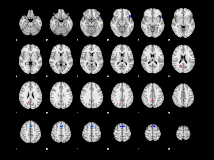
Figure 1 demonstrates the regional associations between brain activation during a verb generation task performed at 12 years of age and exposure to TRAP at 5 years of age. Children with high TRAP exposure levels were associated with regionally specific reductions in brain activation within the left orbital frontal cortex (Figure 2) and the left superior frontal gyrus compared to children with low TRAP exposure.Conclusions
Children with higher exposure to TRAP demonstrate reduced activation within Broca’s area, a region responsible for speech production. The significance of this neuroimaging outcome is being investigated.Acknowledgements
Funding for this project came from the National Institutes of Environmental Health Sciences (NIEHS) R01 ES019890.References
[1] Dockery DW, Ann Epidemiol 2009, 19:257-263
[2] Elder A, et al. Environ Health Perspect 2006, 114:1172-8
[3] Ryan PH, et al. Sci Total Environ 2008, 404:139-147
Figures