4594
An fMRI investigation of the neural efficiency of abstract reasoning as a function of trait anxiety1NMR Research Centre, INMAS, Delhi, India
Synopsis
According to the Attentional Control Theory, trait anxiety has a greater adverse effect on processing efficiency (i.e. performance effectiveness/ effort) than on accuracy. Functional magnetic resonance imaging (fMRI) provides a measure of task-related effort in the form of neural activity elicited during cognitive processing. fMRI was used to assess the neural activation (Blood Oxygen Level Dependent (BOLD) contrast estimates) in a priori regions of interest for a reasoning task. Our results indicate that a compensatory increased neural effort is required by high trait anxious individuals to maintain an equivalent task performance as that of low anxiety individuals.
Introduction
Anxiety is an aversive emotional and motivational state that results in many behavioral and cognitive consequences such as increased distractibility, attentional bias in favor of threat-related information and hyper-responsive amygdala even for unattended threat-related stimuli1. According to the Attentional Control Theory (ACT), processing efficiency, i.e. the effort required for task performance, is affected more by anxiety as compared to performance effectiveness or accuracy of task performance1. The Blood Oxygenation Level Dependent (BOLD) signal during task performance is an index of neural effort that can be related to behavioral performance to derive an estimate of the efficiency of cognitive processing2. In this study, we investigated the reduced processing account of the ACT using a fMRI reasoning task. We investigated whether the neural response of the a priori ROIs to a reasoning task was modulated by variation in the trait anxiety scores in healthy participants.Methods
Twenty two right-handed, healthy and educated (participants (male – 10, female – 12, mean age – 21.09 years, SD – 1.51 years) were recruited from the authors’ home institute. Following the study by Melrose et al. (2007), an fMRI paradigm was designed3. During the reasoning phase, subjects saw three sequential stimuli presented in one row and they had to determine what the fourth picture in the sequence would be. The baseline task was designed to require similar processing in terms of visual encoding, decision process and motor response execution, without the need for reasoning.
The participants were scanned inside a 3 Tesla whole body MRI system (Magnetom Skyra, Siemens, Germany) equipped with a circularly polarized 20 channel matrix head and neck coil and 45 mT/m actively shielded gradient system. Thirty-six axial slices parallel to the bicommissural plane through the fronto-parietal cortex covering the whole brain volume using gradient echo based interleaved EPI sequence (matrix = 64* 64, field of view = 210 mm, TE = 36 ms, TR = 3 s, flip angle = 90˚, slice thickness = 3 mm, voxel size = 3.28 * 3.28 * 3 mm3) were obtained. Two hundred and three sequential image volumes were taken. The baseline (33 sec) and the reasoning task (63 sec) blocks consisted of 4 and 8 stimuli (and response phases) respectively along with an introductory screen (3sec) at the beginning of each condition. The total fMRI acquisition time was 10 minutes and 9 seconds. Stimuli were presented using fMRI hardware from NordicNeuroLab and the subject’s response was monitored with the help of Nordic response device system. The participants completed the Spielberger's State and Trait Anxiety Inventory (STAI)4 and also the Beck Depression Inventory (BDI)5.
fMRI image data were processed with Statistical Parametric Mapping (http://www.fil.ion.ucl.ac.uk/spm) software package implemented in MATLAB R2008a.
A priori regions of interest (ROIs) were determined based on areas activated by reasoning task in a previous study (i.e., bilaterally caudate head, putamen, globus pallidus, thalamus, RLPFC, DLPFC, VLPFC, inferior parietal lobule, superior parietal lobule, middle occipital gyrus (BA 17/18), right postcentral gyrus, left precuneus, right posterior cingulate and right angular gyrus). To sample BOLD activity within these regions, we used 8-mm-radius sphere centered on coordinates from Melrose et al. (2007)3. BOLD contrast estimates were then extracted from these ROIs using MarsBaR toolbox of SPM (http://marsbar.sourceforge.net/). Partial correlation analysis between BOLD contrast estimates in all the ROIs and trait anxiety scores of the subjects (n = 22) was also carried out to detect regions where BOLD contrast estimates correlated with the trait anxiety. Age, sex, BDI scores and STAI-Y1 scores were taken as covariates.
Results
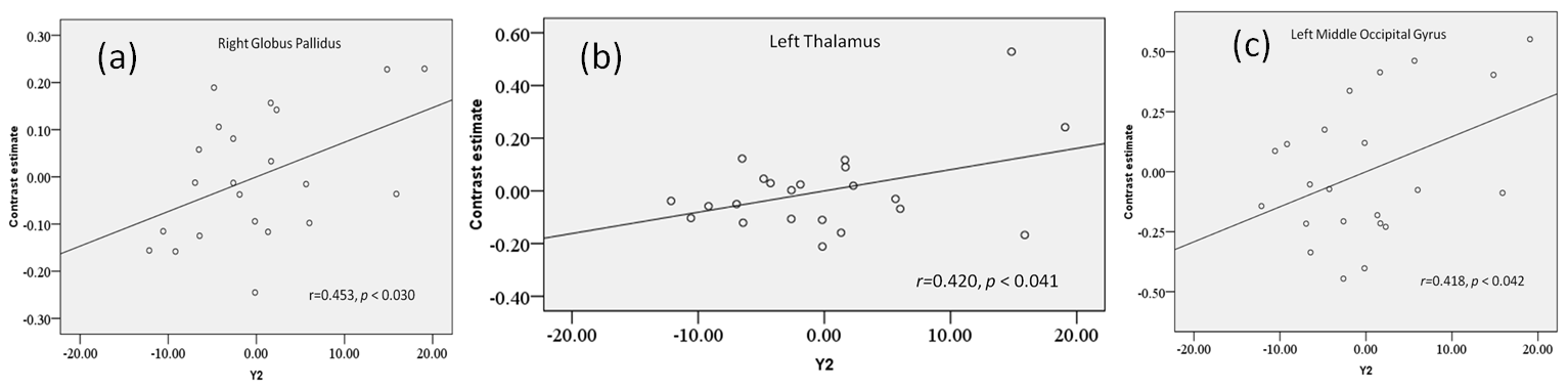
Descriptive statistics for the self-report measures are as follows: state anxiety score (STAI-Y1) = 37.41 ± 10.8, trait-anxiety score (STAI-Y2) = 41.32 ± 9.40, BDI = 9.23 ± 2.41. A significant positive correlation between trait anxiety scores and contrast estimates was obtained in right globus pallidus (n = 22, r=0.453, p < 0.030), left thalamus (n = 22, r=0.420, p < 0.041) and left middle occipital gyrus (n = 22, r=0.418, p < 0.042) (Figure 1 a, b, c). A trend towards correlation was obtained in left globus pallidus (n = 22, r=0.351, p < 0.076) and left DLPFC (n = 22, r=0.380, p < 0.060) (Figure1).Discussion
Correlation analysis findings might suggest a possibly stronger engagement of sensory pathways as well as visual encoding, analysis and attention systems in high trait anxiety individuals thereby providing a plausible physiological mechanism by which they might exhibit comparable performance as that of low trait anxious individuals. The increased activity might sub serve a compensatory mechanism for equivalent task performance in high trait anxious individualsAcknowledgements
No acknowledgement found.References
1. Eysenck MW, Derakshan N, Santos R, et al. Anxiety and cognitive performance: attentional control theory. Emotion 2007; 7(2): 336-353.
2. Basten U, Stelzel C, Fiebach CJ. Trait anxiety modulates the neural efficiency of inhibitory control. Journal of Cognitive Neuroscience 2011; 23(10): 3132-3145.
3. Melrose RJ, Poulin RM, Stern CE. An fMRI investigation of the role of the basal ganglia in reasoning. Brain Research 2007; 1142: 146 – 158.
4. Spielberger CD. Manual for the state-trait anxiety inventory. Consulting Psychologists Press, Palo Alto, CA 1983.
5. Beck AT, Steer RA, Brown GK. Beck depression inventory: Manual. 2nd ed. The Psychological Corporation, San Antonio1996.
Figures