4593
Abnormally functional connectivity density and network pattern in lifelong premature ejaculation patient: A resting-state fMRI study1Department of Radiology, Affiliated Drum Tower Hospital of Nanjing University Medical School, Nanjing, Jiangsu, China, NanJing, People's Republic of China, 2Department of Andrology, Affiliated Drum Tower Hospital of Nanjing University Medical School, Nanjing, Jiangsu, China, NanJing, People's Republic of China, 3Department of Urology, The First Affiliated Hospital of Nanjing Medical University, Nanjing, Jiangsu, China., 4Department of Radiology, The Affiliated Yancheng Hospital of Southeast University Medical College, Yancheng, Jiangsu, China, NanJing, People's Republic of China, 5Philips Healthcare, Shanghai, People's Republic of China, People's Republic of China, 6Philips Healthcare, HongKong, People's Republic of China, People's Republic of China, 7Department of Radiology, Affiliated Drum Tower Hospital of Nanjing University Medical School, Nanjing, China, NanJing, People's Republic of China, 8Department of Radiology, ffiliated Drum Tower Hospital of Nanjing University Medical School, Nanjing, Jiangsu, China., NanJing, People's Republic of China
Synopsis
Premature ejaculation (PE) is considered the alteration in dopaminergic reward system. Functional connectivity density along with network pattern methodology provides an opportunity to assess the functional integrity of brain activity in implicated circuits and it was applied to analysis the inter-group difference in SFCD and LFCD. Network constructing and graph theoretical were used to compare the network property. In conclusion, this study demonstrated the weaken connection inner brain areas and strengthened connection inter brain areas in dopaminergic reward system along with the functional network pattern has changed in the lifelong PE patient for the first time.
PURPOSE
It is hypothesized that premature ejaculation (PE) is considered the alteration in dopaminergic reward system1. However, findings of functional abnormalities using MRI have been inconsistent. Functional connectivity density along with network pattern methodology provides an opportunity to assess the functional integrity of brain activity in implicated circuits.METHODS
The study population was comprised of 20 right-handed individuals with lifelong PE (age: 27.95±4.52, male) and 15 age- and gender-matched normal control (age: 27.87±3.78, male). All participants underwent a 3.0-tesla resting state and T1 weighted fMRI scan at Philips 3T MR Scanner(Achieva TX, Best, the Netherlands). Functional connectivity density (FCD) was applied to analysis the inter-group difference in SFCD and LFCD. 42 functional connectivity hubs from literature as ROIs to perform ROI-based analysis. Network constructing and graph theoretical were used to compare the network property. We further analyzed that the correlation between IELT score with FCD and network property by Pearson correlation.RESULTS
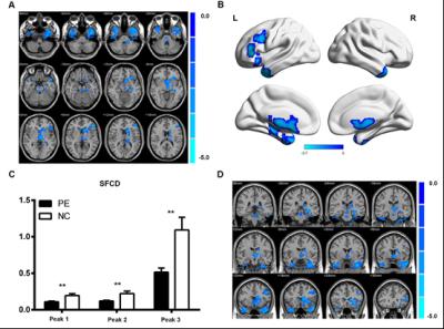
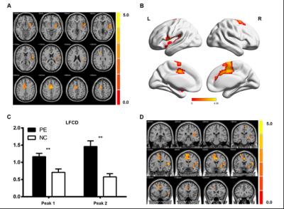
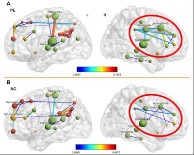
Using FCD and ROI-based analysis, we found significant decreased SFCD in bilateral middle temporal gyrus, bilateral fusiform, the left orbitofrontal cortex (OFC), left nucleus accumbens and left fusiform, left caudate and left thalamus (p<0.05, AlphaSim corrected). Increased long-rang FCD are found in left insula, left heschl, left putamen, bilateral precuneus, bilateral support moto area, middle cingulate cortex (MCC), anterior cingulate cortex (ACC). No increased SFCD and decreased LFCD difference are found between the two groups. Graph theoretical analysis found altered network pattern and reinforced connection among these nodes: right ACC, right inferior frontal, bilateral nucleus accumbens, right insula, left heschl, right MCC and right inferior temporal. Also, the degree of right ACC, right inferior frontal and right MCC has increased in PE patients. The IELT has positive correlation with SFCD and negative correlation with LFCD and node degree.DISCUSSION
To the best of our knowledge, this is the first study applying FCD mapping to explore aberrant rsFC in lifelong PE. Our findings showed that PE patients had significantly enhanced long-range FCD in left insula, left heschl, left putamen, bilateral support moto area, middle cingulum gyrus, right anterior cingulum gyrus and decreased short-range FCD in the bilateral middle temporal gyrus, bilateral fusiform, left dorsolateral prefrontal cortex (DLPFC), left caudate and left thalamus, which reflects the disrupted inter-regional FC in brain. Further graph theoretical and network-based statistical analyses found the global efficiency, local efficiency, clustering coefficient and node degree are increased in PE patients. Moreover, the network strength has reinforced between right ACC, right inferior frontal, bilateral nucleus accumbens, right insula, left heschl, right MCC and right inferior temporal. Also, the degree of right ACC, right inferior frontal and right MCC has increased in PE patients. This study provides new insight into the pathophysiological mechanisms of premature ejaculation. 2,3, which mainly includes subcortical structures such as the ventral tegmental area, substantia nigra, basal ganglia or amygdala, and prefrontal areas such as the orbitofrontal cortex (OFC), medial prefrontal cortex (PFC), and anterior cingulate cortex (ACC)4,5. The weakened local association in bilateral middle temporal gyrus, bilateral fusiform, the left orbitofrontal cortex (OFC), left caudate and left thalamus are observed in our data. Previous studies have proposed a theory that reinforcement sensitivity theory (RST), which proposes the existence of a neurobehavioral system involved in the processing of appetitive stimuli2. The primary function of this system is to bring together the individual with biological rewards such as sex and food. The biological substrate of this system is thought to comprise brain areas belonging to the dopaminergic reward system.
In conclusion, this study demonstrated the weaken connection inner brain areas and strengthened connection inter brain areas in dopaminergic reward system along with the functional network pattern has changed in the lifelong PE patient for the first time. We hoped that these findings would enhance understandings of PE and provide a new approach in future studies and new therapy development.
Acknowledgements
No acknowledgement found.References
1 Laumann, E. O. et al. Sexual problems among women and men aged 40-80 y: prevalence and correlates identified in the Global Study of Sexual Attitudes and Behaviors. International journal of impotence research 17, 39-57, doi:10.1038/sj.ijir.3901250 (2005).
2 Costumero, V. et al. Reward sensitivity is associated with brain activity during erotic stimulus processing. PloS one 8, e66940, doi:10.1371/journal.pone.0066940 (2013).
3 Pickering, A. D. & Gray, J. A. The neuroscience of personality. Handbook of personality: Theory and research 2, 277-299 (1999).
4 Haber, S. N. & Knutson, B. The reward circuit: linking primate anatomy and human imaging. Neuropsychopharmacology 35, 4-26 (2010).
5 Ikemoto, S. Dopamine reward circuitry: two projection systems from the ventral midbrain to the nucleus accumbens–olfactory tubercle complex. Brain research reviews 56, 27-78 (2007).
Figures


