4577
The Sensorimotor Network Dysfunction in Migraineurs WithoutAura: A Resting-state fMRI study1Shanghai Key Laboratory of Magnetic Resonance and Department of Physics, School of Physics and Materials Science, East China Normal University, Shanghai, People's Republic of China, 2Department of Neurology and Jiuyuan Municipal Stroke Center, Shanghai Ninth People's Hospital, Shanghai Jiao Tong University School of Medicine, Shanghai, People's Republic of China
Synopsis
In current study, we analyzed resting-state fMRI data for the first time to evaluate the dysfunction of the sensorimotor network in migraineurs without aura by applying regional homogeneity (ReHo), amplitudes of low-frequency fluctuation (ALFF) and degree centrality (DC) analysis methods. The ReHo, DC and ALFF values were decreased in the S1 and PMC indicating the sensorimotor network dysfunction in migraineurs without aura. These changes may result in disruption of discrimination of sensory features of pain, thereby affecting nociception pathways. Non-invasive brain stimulation could be applied to sensorimotor network to modulate headache pain in future therapies.
Introduction:
Migraine is a common and refractory neurological disorder combining nausea, vomiting, and hypersensitivities to visual, auditory, olfactory and somatosensory stimuli1. In the past decade, functional magnetic resonance imaging (fMRI) is used to investigate the pathophysiology mechanism of migraine and has led to an evolution in knowledge about migraine. However, the dysfunction of the sensorimotor network in migraineurs has not been well clarified.Materials and Methods:
The rest state functional MRI images were acquired using gradient-echo EPI pulse sequence with the following parameters: repetition time = 2000 ms, echo time = 30 ms,number of slices = 33, total volumes = 210. In present study, we evaluated the dysfunction of sensorimotor network in 30 migraineurs without aura (8 males, 22 females)by combining regional homogeneity (ReHo), amplitudes of low-frequency fluctuation (ALFF) and degree centrality (DC) analysis methods based on resting state fMRI comparing with 31 controls (9 males, 22 females). The seed-based functional connectivity (FC) analysis was used to investigate whether the dysfunction areas within sensorimotor network exhibit abnormal FC with other brain areas.Results:
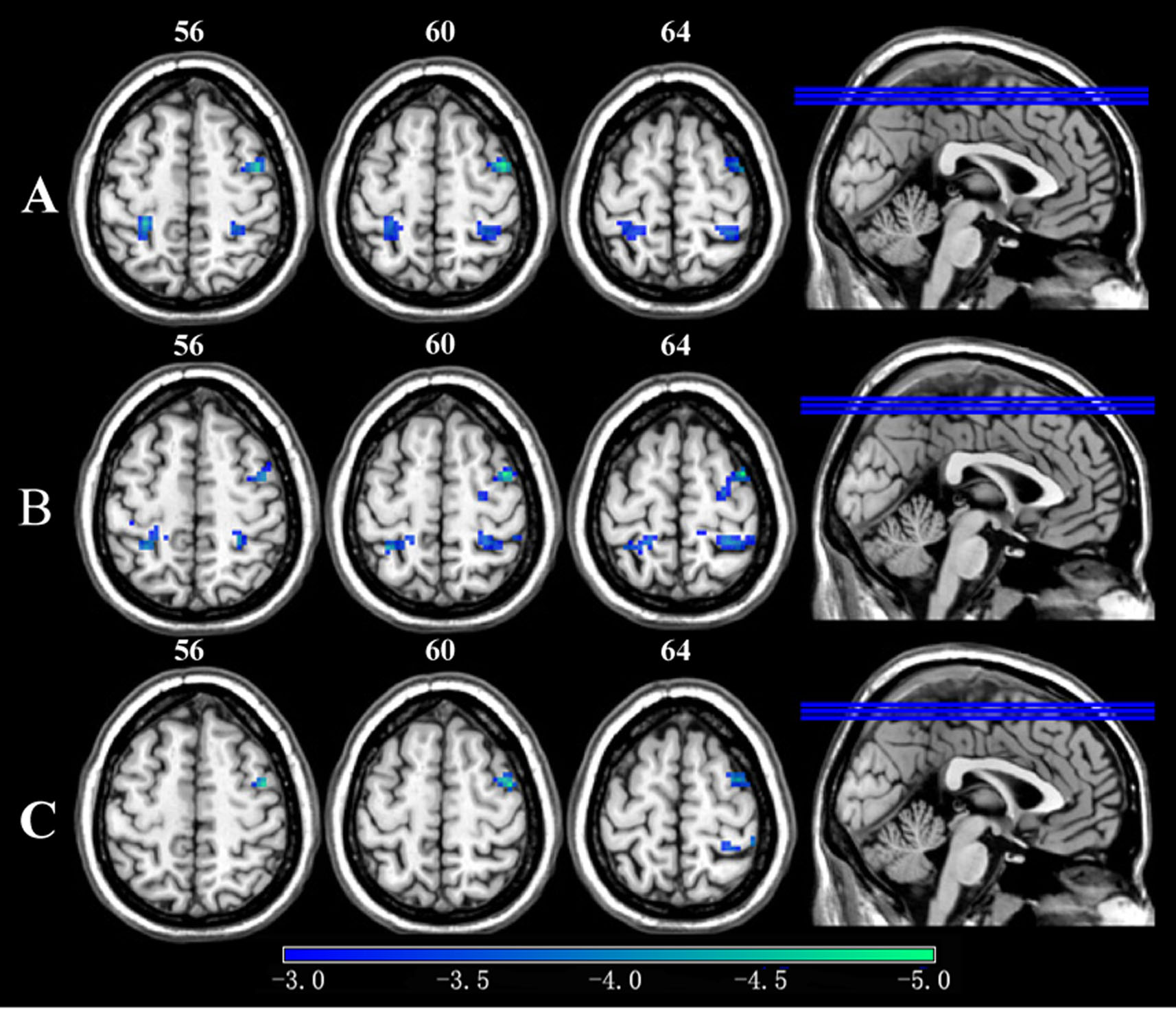
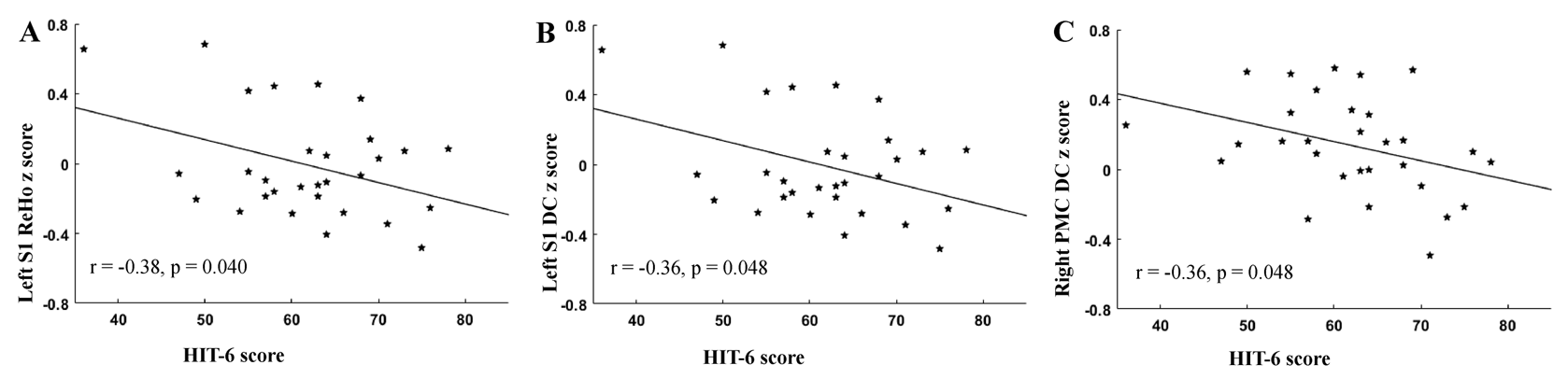
Compared with the controls, the migraineurs without aura exhibited significantly decreased ReHO, ALFF and DC values in the primary somatosensory cortex (S1) and right primary motor cortex (PMC) (Figure 1).We found that the ReHo z score and the DC z score of left S1 and the DC z score of right PMC were negatively correlated with Headache Impact Test (HIT-6) score (Figure 2). In our study, compared with controls, the migraineurs also showed weaker FC between the S1 and many other many areas , such as right temporal lobe, inferior and superior parietal lobules, ACC, insular cortex, and pons, which are involved in pain discrimination and/or pain processing.Discussion:
We proposed that the dysfunction of S1 and PMC and decreased FC between S1 and brain areas in migraineurs without aura may result in disruption of discrimination of sensory features of pain and would affect trigemino-thalamo-cortical pathway in migraine, which maybe paly play an important role in somatosensory hypersensitivity and central sensitization 2,3. In future therapies, non-invasive brain stimulation, such as transcranial magnetic or direct current stimulation, can be applied to sensorimotor network (especially S1 and PMC), may potentially modulate chronic pain.Acknowledgements
This work was supported by grants from the National Natural Science Foundation of China (Nos. 81571658 and 81201082 to X. X. Du)References
1. Schwedt TJ, Chiang CC, et al. Functional MRI of migraine. The Lancet. Neurology. 2015;14(1):81-91.
2. Oshiro Y, Quevedo AS, et al. Brain mechanisms supporting spatial discrimination of pain. The Journal of neuroscience : the official journal of the Society for Neuroscience. 2007;27(13):3388-3394.
3. Burstein R, Noseda R,et al. Migraine: multiple processes, complex pathophysiology. The Journal of neuroscience : the official journal of the Society for Neuroscience. 2015;35(17):6619-6629.
*Correspondence to: Xiaoxia Du. E-mail:xxdu@phy.ecnu.edu.cn.
Figures