4547
Improvement of dynamic improved motion-sensitized driven-equilibrium steady-state free precession (dynamic iMSDE SSFP) to visualize the irregular motion of cerebrospinal fluid1Department of Radiology, Tokai University Hospital, Isehara, Japan, 2Department of Radiology, Tokai University School of Medicine, Isehara, Japan, 3Department of Neurosurgery, Tokai University School of Medicine, Isehara, Japan, 4Course of Electrical and Electronic Engineering, Graduate School of Engineering, Tokai University, Hiratsuka, Japan, 5Healthcare, Philips Electronics Japan Ltd, Shinagawa, Japan, 6Department of Radiology, Tokai University Hospital, Hachiouji, Hachiouji, Japan
Synopsis
We reported a new technique to visualize the irregular motion of cerebrospinal fluid (CSF) by using dynamic improved motion-sensitized driven-equilibrium steady-state free precession (dynamic iMSDE SSFP). The purpose of this study was to optimize the sequence parameters of dynamic iMSDE SSFP. As a result, the slow and irregular CSF motions were sensitively detected using the following parameters: T2prepTE: 30 ms, dynamic interval: 700 ms, flow VENC: 1 cm/s and the directions of MSG: 3axes. Therefore, optimized dynamic iMSDE SSFP is suggested to contribute to the diagnosis of various diseases in the CSF space.
INTRODUCTION
We reported a new technique to visualize the irregular motion of cerebrospinal fluid (CSF) by using dynamic improved motion-sensitized driven-equilibrium steady-state free precession (dynamic iMSDE SSFP). [1] To visualize CSF motion and accentuate its features, a selected image (tenth image) with motion-sensitized gradient (MSG)-off was subtracted from those with MSG-on as shown in Figure 1. This method has advantages in no requirement of pulse triggering or complex post-processing of images, and allows visualization of CSF motion in a short period of time in selected whole imaging planes. However, as shown in Figure 2, T2prepTE, dynamic interval, for flow VENC and directions of MSG has not still been optimized to detect CSF motions. The purpose of this study was to optimize the sequence parameters of dynamic iMSDE SSFP.METHODS
The equipment we used was a 1.5T clinical scanner (Achieva nova dual, Philips, Best, the Netherlands) with either a 32ch SENSE torso cardiac coil (phantom study) or a 6ch SENSE head coil (volunteer study). The basic parameters for dynamic iMSDE SSFP were as follows: 2D balanced TFE with iMSDE, FOV of 256×256 mm2, matrix of 192×256, TR/TE: 3/1.51 ms, slice thickness of 10 mm, flip angle of 90°, reduction factor: 2, turbo field echo factor: 90, NSA:1, and dynamic scan: 30 (MSG-off : 15, MSG-on : 15). Phantom study: The flow phantom was made with superabsorbent polymer and tubes with steady flow. We investigated the optimal T2prepTE and dynamic interval. The signal intensity of flowing water in the tube was measured with varying T2prepTE (15, 30, 100, and 200 ms) and dynamic interval (0, 300, 700, and 1700 ms) in longitudinal section. Differences of signal intensity for each parameter were assessed by using one-way repeated-measures analysis of variance (ANOVA) with multiple- comparison Dunn's test. Volunteer study: five healthy volunteers (age range, 25–45 years) were included; written informed consent was obtained from all volunteers. We examined the flow VENC and directions of MSG in the mid-sagittal cross-section in the volunteers. Visual assessment was performed using a 5-point scale by five radiologists. The criteria of the 5-point scale were: 1 (unsatisfactory), 2 (poor image), 3 (fair image), 4 (good image), and 5 (excellent image). Differences of the scores for each parameter were assessed by using the Friedman’s test and multiple-comparison Holm's test.RESULTS
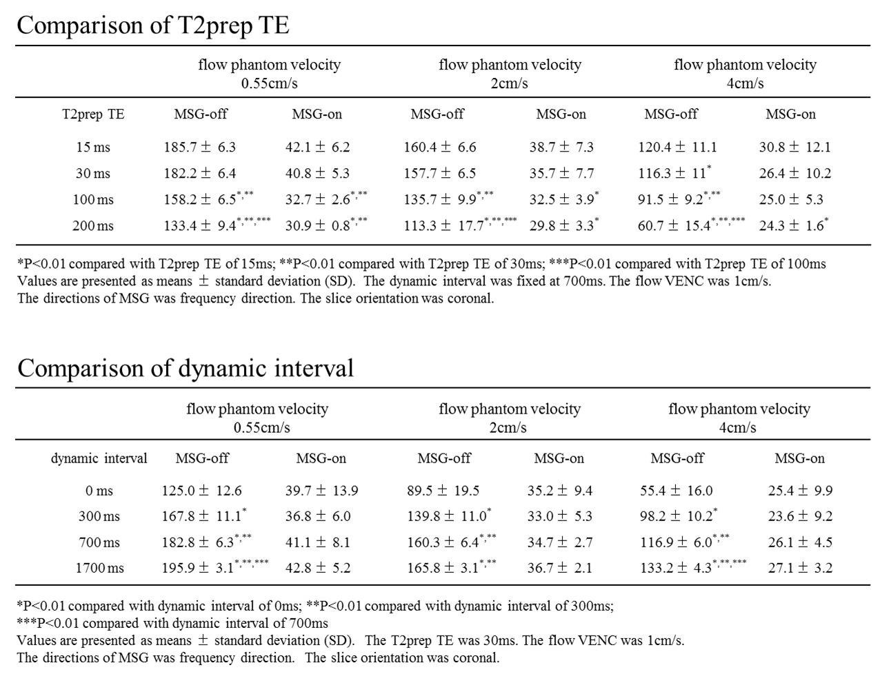
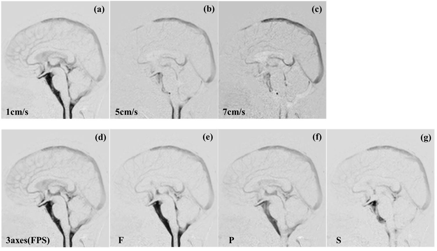
Phantom study, T2prepTE: signal intensity of MSG-off decreased by extending the T2prep TE, but that of MSG-on slightly decreased. Dynamic interval: signal intensity of MSG-off increased by extending the dynamic interval, but that of MSG-on did not show significant changes. These results were similar in all flow rates. (Table1) Volunteers study: As shown in Figure 3, dynamic iMSDE SSFP with flow VENC: 1cm/s, directions of MSG: 3 axes (FPS : frequency, phase, and slice directions) achieved significantly higher score. (P <0.01) In the case of 3-axis observation, visibility of CSF flow with 1-cm/s VENC was clearer than that with 5 or 7cm/s as shown in Figure 4(a-c). As also appreciated from (d-g), the 3-axes (FPS) image revealed the CSF flow in the lateral ventricle, third ventricle, fourth ventricle, ventral surface of the brain stem, and cisterna magna with higher contrast than the single-axis images.DISCUSSION
Phantom study: The technique requires signal-intensity differences between MSG-off and MSG-on for subtraction. More signal-intensity differences were obtained by shortest T2perpTE and extension of dynamic interval. However, the shortest T2prepTE limit the use of flow VENC, and long dynamic interval decreased the temporal resolution. Therefore, T2prepTE: 30 ms and dynamic interval: 700 ms were the optimum. Volunteers study: Detection of slow and irregular motion in CSF required a small flow VENC and 3 axes directions of MSG. Optimal sequence parameters were as follows; T2prep TE: 30 ms, dynamic interval: 700 ms, flow VENC: 1 cm/s, directions of MSG: 3 axes, and the temporal resolution, one second.CONCLUSION
Optimized dynamic iMSDE SSPF can sensitively detect the slow and irregular CSF motions. Therefore, this technique is suggested to contribute to the diagnosis of various diseases in the CSF space.Acknowledgements
No acknowledgement found.References
[1] Horie T, et al. Magnetic resonance imaging technique for visualization of irregular cerebrospinal fluid motion in the ventricular system and subarachnoid space. World Neurosurg. 2016 Jul 26. pii: S1878-8750(16)30590-3.Figures
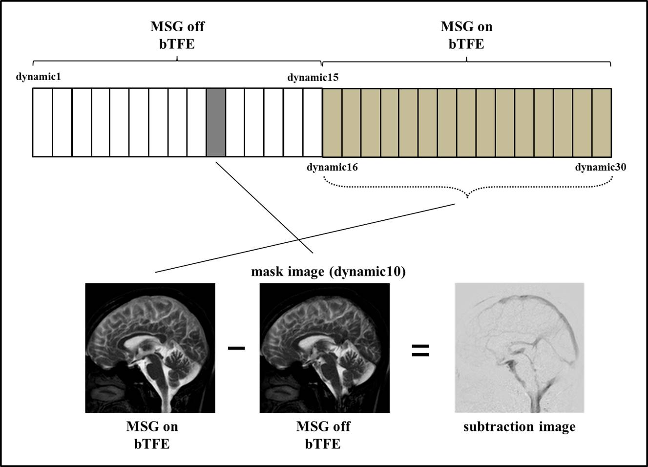
Figure 1 Schematic diagram of the proposed dynamic iMSDE SSFP design. Balanced turbo field-echo (bTFE) acquisitions with and without MSG preparation are dynamically obtained 15 times, each at an identical slice.
To create motion-sensitized contrast, subtraction of the MSG-on and -off images was performed. The tenth dynamic image without MSG was selected and used for the subtraction.
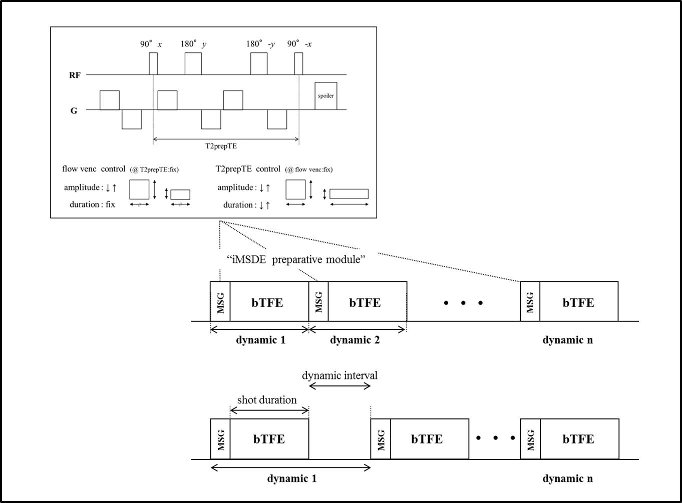
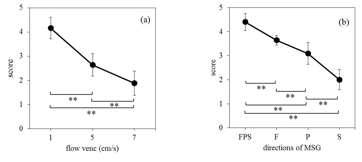
Figure 3 Visual assessment of the volunteers.
(a) Comparison of the flow VENC. Flow VENC 1cm/s shows significantly higher scores than 5cm/s and 7cm/s. The directions of MSG were 3 axes (FPS : frequency, phase, and slice direction). * P<0.01
(b) Comparison of the directions of MSG. 3 axes (FPS) was shows significantly higher scores than F, P and S. The directions of MSG was 3 axes (FPS : frequency, phase, and slice direction).
The flow VENC was 1cm/s. * P<0.01