4535
Longitudinal changes in cortical thickness in collegiate high contact sports1Stanford University, Stanford, CA, United States
Synopsis
There is emerging evidence that cumulative sports-related concussions may lead to long-term neurological abnormalities. The extent to which changes are occurring in collegiate athletes is still undetermined, and the progression of these changes is poorly understood. Cross sectional studies with small cohorts have found that concussions maybe be associated with cortical thinning in young football players (1,2). We analyze the longitudinal changes occurring in high vs. low contact sports over the course of 3-4 years.
Target audience
Scientists/MR physicists/Neurologist/Radiologists with interests in traumatic brain injury and longitudinal morphometric analysisPurpose
Our objective is to perform a longitudinal analysis of the effect of concussive and sub-concussive traumatic brain injury in high-contact sports on cortical integrity, specifically cortical thickness estimates.Methods
Players and Image acquisition
A total of 255 brains scans were acquired from 62 high-contact (football) players and 24 low-contact (volleyball) players over the course of 3-4 years, in accordance with IRB and HIPAA. Players were scanned at the start of each season, within 24-96 hours of a concussion, and before finishing college, with each player having 1-6 time-points based on study enrollment timing. Using a 3T scanner (GE, Milwaukee, WI, USA) and an 8-channel head coil, we acquired multiple sequences, and here report results on the whole-brain structural T1-weighted images (FSPGR BRAVO, 0.93x0.93x1mm, 5 minutes). In addition to imaging, all high-impact players were evaluated using the standardized sport concussion assessment tool II (SCAT II) at baseline and after concussion.
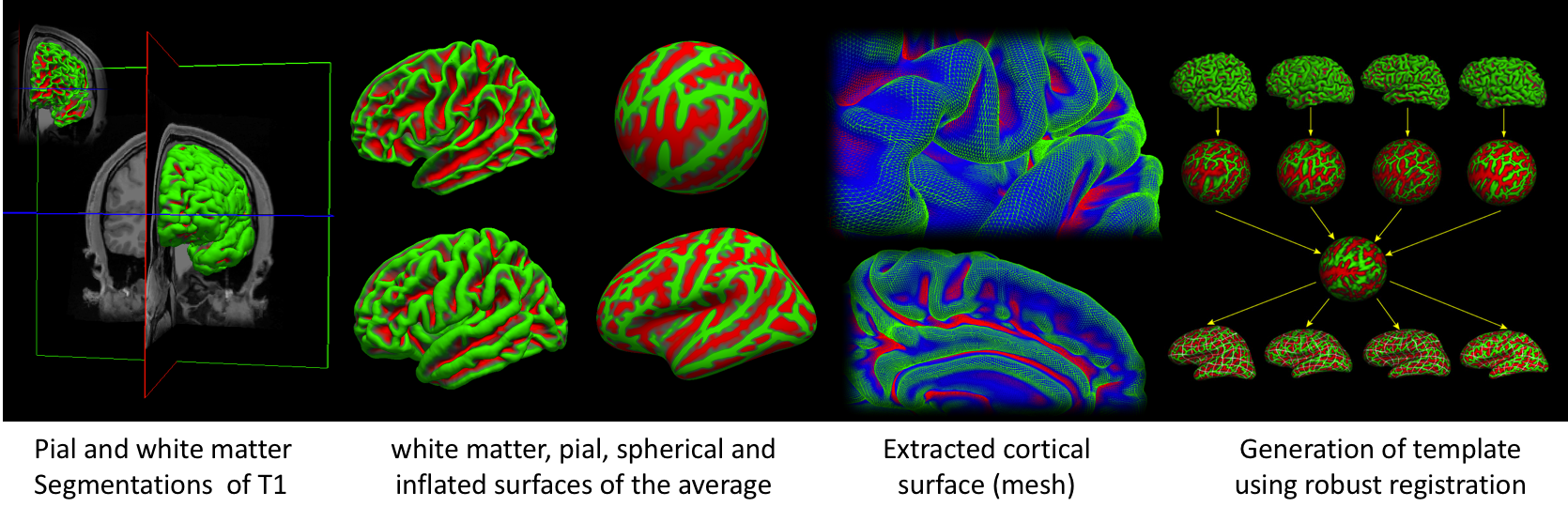
Freesurfer longitudinal pipeline and cortical thickness estimation
The longitudinal processing stream in Freesurfer (v5.3) was used: this first generates an unbiased, within subject template by iteratively aligning all input images to a median image (3). This “base” was then iteratively edited manually, (by two successive blinded raters) to ensure accurate segmentation, by altering the scalp mask and placement of control points. Then, we initialized the processing of a new data set in a longitudinal series of the same subject with his common template (Figure 1), which are also subject to the same blinded manual editing steps. Cortical thickness was measured as the distance between the gray–white matter boundary and the pial surface at each point of the cortical surface (4). Cortical thickness estimates were resampled to an averaged surface template and smoothed with a 30mm full-width half-maximum Gaussian kernel.
Statistical analysis
We employed a mass-univariate, spatiotemporal linear mixed effects (LME) model (5) in MATLAB (2014b, Mathworks) to address the local spatial correlations as well as the temporal correlations among repeated measures of the same subject. To determine regionally specific differences in cortical thickness atrophy rate over time among high and low contact sports, our model included time (days) at scan, age at baseline, sport (football or volleyball) and sport-time interaction as fixed effects, with a random intercept. Including time as a random effect did not improve the fit of the model. Multiple comparison correction was performed using false discovery rate (FDR) with an alpha of 0.05 for each vertex of both hemispheres.
Results and Discussion
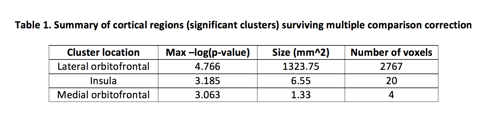
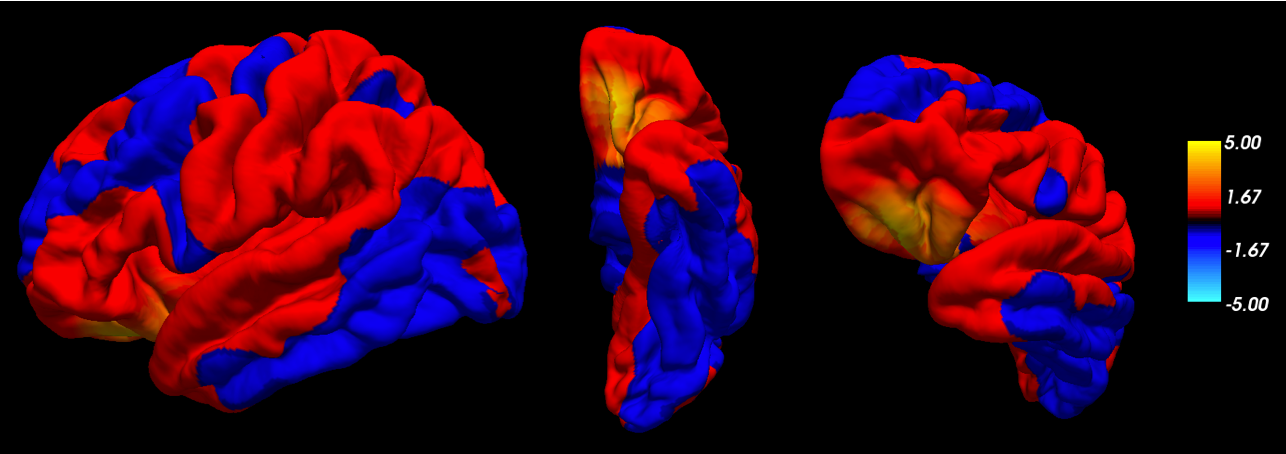
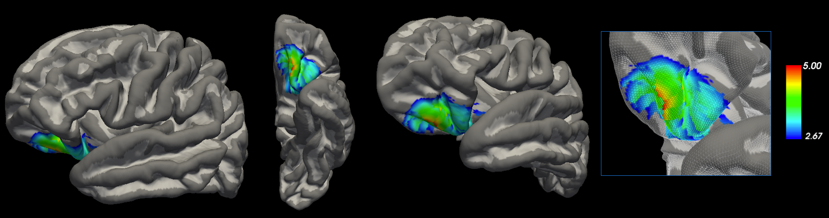
Figure 2 demonstrates significant, uncorrected regions (red) with altered atrophy rates between high contact sports vs. low contact sports (interaction between sport and time). Three significant clusters within the left hemisphere highlighting differences in cortical thinning between the two groups (type of sport) survived multiple comparison correction (Figure 3). When filtering these clusters using their area (mm2) only one adequately large cluster, located in the left lateral orbitofrontal region of the brain, survived (Table 1). No clusters survived multiple comparison correction in the right hemisphere.
Future work will assess if cortical atrophy rates differ between football players with a history of concussion and those without reported concussion, as compared to controls. The left lateral orbitofrontal region is susceptible to segmentation errors; thus future work will also include further validation of parcellation accuracy in the region.
Conclusion
Using a spatiotemporal linear mixed effects model, we found a significant interaction between time and sport in the left lateral ortbitofrontal region. This region may be susceptible to the effects of high-contact sports.Acknowledgements
This work was funded by the radiological society of north america (RSNA) and general electric (GE).References
[1] Meier et al., Journal of Neurotrauma 2016
[2] Tremblay et al., Clinical Neurophysiology 2014
[3] Reuter et al., Neuroimage 2012
[4] Reuter et al., Neuroimage 2013
[5] Bernal-Rusiel et al., Neuroimage 2013
Figures