4532
Measurements of Microstructural Changes after Sildenafil Treatment of Experimental Traumatic Brain Injury in Rats using Diffusion Tensor Imaging1Center for Neuroscience and Regenerative Medicine, Henry Jackson Foundation, Bethesda, MD, United States, 2Radiology and Radiological Sciences, Uniformed Services University of Health Sciences, Bethesda, MD, United States, 3Center for Neuroscience and Regenerative Medicine, Uniformed Services University of Health Sciences, Bethesda, MD, United States, 4Neurology, Uniformed Services University of Health Sciences, Bethesda, MD, United States
Synopsis
The long-term dynamic response of microstructural changes in rat brain to the administration of sildenafil after traumatic brain injury (TBI) using non-invasive MRI techniques have been investigated. Our results demonstrate that the treatment of diffuse traumatic brain injury with sildenafil reverses several changes in brain microstructure at 30 days post injury. Diffusion tensor imaging (DTI) data suggests that sildenafil treatment improves white matter reorganization after TBI in rats compared with saline treatment.
Introduction
Traumatic brain injury (TBI) is a devastating disease with a variety of cognitive and motor function deficits that can manifest for several minutes to years after the initial impact, making it an important public health problem1. TBI produces a primary insult, which may trigger widespread brain damage regardless of the original site of injury2. Thus, patients with TBI may suffer from axonal degeneration and generalized cerebral atrophy associated with poor neurological functions3. Currently, there is no effective clinical treatment that can repair biostructural damage to the neurons and prevent or reduce secondary pathological processes4. Experimental pharmacological treatments of TBI that promote brain remodeling have shown promising results in improving functional recovery after TBI in animals. Administration of sildenafil potentiates nitric oxide signaling by inhibiting cGMP breakdown increasing brain levels of cGMP5. Elevated cGMP levels in cerebral tissues may enhance the improvement of functional outcome, angiogenesis and synaptogenesis after brain injury5. The therapeutic effects of treatment of TBI can be dynamically monitored and longitudinally investigated using magnetic resonance imaging (MRI).Materials and Methods
Male Sprague-Dawley (SD) rats (200-300g) subjected to the experimental traumatic brain injury model, fluid percussion injury (FPI), were randomly assigned to sildenafil (n=7) or saline (n=8) treatment groups. Sildenafil was administered at a dose of 10 mg/kg subcutaneously in the treated group 1 hour after TBI and daily for an additional 6 days. Rats in the control group were treated with the same volume of saline. In vivo MRI was performed on all the rats prior to the injury (baseline), as well as 1, 7, and 30 days after the injury. All rats were sacrificed at 30 days after injury for histological analysis.MRI Protocol
MR measurements including multi-echo T2 (TR=6300ms, TE=20, 60, 100 and 140ms; FOV=25.6mm and thickness=1mm; number of slices=15), 3D multi gradient echo for T2* (TR=45ms,TE=4, 8, 12, 16, 20, 24 and 28ms; FOV=25.6mm; thickness=1mm; number of slices=11), cerebral blood flow (CBF) measurement with the arterial spin labeling (cASL) technique (TR=7000ms,TE=20ms; FOV=25.6mm and thickness=1mm; number of slices=5), and Diffusion Tensor Imaging (DTI) (TR=4000ms,TE=24ms; FOV=2.56mm; 12 directions thickness=1mm; number of slices=15 ) were performed on the 7 Tesla 20-cm horizontal bore Bruker BioSpec system (Bruker Biospin, Billerica, MA) equipped with an 86mm quadrature transmit coil, and a dedicated phased array head receiver coil. Anesthesia was delivered using isofluorane (2%) and nitrogen/oxygen mixture (70%/30%). The body temperature was maintained at 37°C, and the respiration rate, heart rate, and blood O2 level were monitored during MRI scans. DTI data was processed using Tolerably Obsessive Registration and Tensor Optimization Indolent Software Ensemble (TORTOISE) software6.Results and Discussion
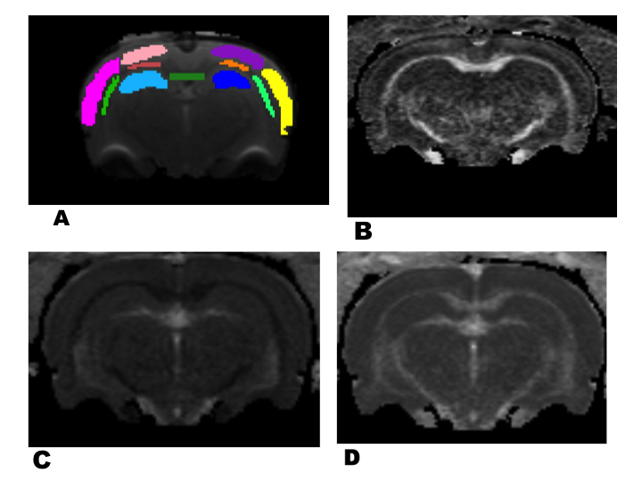
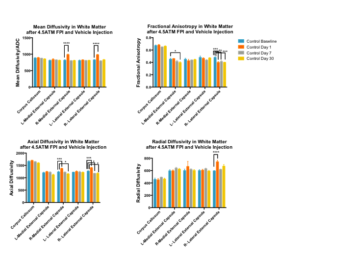
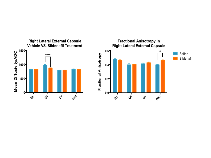
Analysis of diffusion tensor imaging was completed using 5 regions of interest (ROIs) for white matter: corpus callosum, the left and right medial segments of the external capsule, and the left and right lateral segments of the external capsule; as illustrated in Figure 1A. The longitudinal changes in DTI computed mean diffusivity (MD), fractional anisotropy (FA), axial diffusivity (AD), and radial diffusivity (RD) for ROIs in the selected white matter regions are shown in Figure 2. The MD was significantly increased in the medial and lateral segments of the ipsilateral external capsule at one day after injury. The FA values for the ipsilateral segment of the external capsule decreased at day 1, 7 and 30 after injury compared to the baseline measurements. Sildenafil treatment reduces the increase in MD observed at day 1 post injury for lateral segments of the ipsilateral external capsule. Conversely, the FA values at the same region increased by day 30 after injury in the sildenafil treated group as shown in Figure 3.Conclusions
Our results reveal that DTI is able to detect subtle microstructural changes induced by experimental traumatic brain injury in a location distal from the lesion site 30 days after the injury. The DTI data suggest that sildenafil treatment improves white matter reorganization at the external capsule at 4 weeks after TBI in rats compared with matched saline treated controls. The spatiotemporal information DTI revealed is extremely valuable in determining the progression of structural cerebral damage after injury. Further studies are required to better understand how sildenafil alters the microstructural abnormalities and change the pathophysiological course of disease.Acknowledgements
Supported by the U.S. Department of Defense in the Center for Neuroscience and Regenerative Medicine (CNRM), with technical support from the CNRM Translational Imaging Facility and Pre-clinical Core, and Uniformed Services University of Health Sciences Grant G192JD13References
1. Stocchetti N, Paternò R, Citerio G, Beretta L, Colombo A Traumatic brain injury in an aging population J Neurotrauma 2012; 29:1119-1125
2. Masel BE, DeWitt DS. Traumatic brain injury: a disease process, not an event. J Neurotrauma 2010; 27:1529-1540
3. Sidaros A, Skimminge A, Liptrot MG, Sidaros K, Engberg AW, Herning M, Paulson OB, Jernigan TL, Rostrup E. Long-term global and regional brain volume changes following severe traumatic brain injury: a longitudinal study with clinical correlates Neuroimage 2009; 44:1-8
4. Guang Liang Ding , Michael Chopp , David J. Poulsen , Lian Li , Changsheng Qu , Qingjiang Li, Siamak P. Nejad-Davarani , John S. Budaj , Hongtao Wu , Asim Mahmood, Quan Jiang. MRI of Neuronal Recovery after Low-Dose Methamphetamine Treatment of Traumatic Brain Injury in Rats. PLoS ONE 2013; 8(4): e61241
5. Zhang L, Zhang RL, Wang Y, Zhang C, Zhang ZG, Meng H, Chopp M. Functional recovery in aged and young rats after embolic stroke: treatment with a phosphodiesterase type 5 inhibitor. Stroke.2005;36:847–852
6. Pierpaoli C. et al TORTOISE: an integrated software package for processing of diffusion MRI data, 2010 ISMRM annual meeting.
Figures