4530
Characterizing White Matter Microstructural Changes After Mild Traumatic Brain Injury Based On Diffusion White Matter Tract Integrity And Shannon Entropy1Center for Advanced Imaging Innovation and Research (CAI2R), Department of Radiology, New York University School of Medicine, New York, NY, United States, 2Bernard and Irene Schwartz Center for Biomedical Imaging, Department of Radiology, New York University School of Medicine, New York, NY, United States, 3Department of Neurology, New York University Langone Medical Center, New York, NY, United States, 4Department of Rehabilitation Medicine, New York University Langone Medical Center, New York, NY, United States
Synopsis
Mild traumatic brain injury (MTBI) is a growing public health problem and some patients may suffer from long-term symptoms. This study shows that there are both microstructural changes as well as regional textural changes after MTBI affecting the corpus callosum within 4 weeks of injury. We demonstrate the potential for compartment specific white matter tract integrity (WMTI) metrics such as tortuosity of the extra-axonal space (a marker of misalignment of fibers or demyelination), and Shannon entropy (reflecting complexity or uncertainty) to be useful as early biomarkers of MTBI-related WM injury.
PURPOSE
Mild traumatic brain injury (MTBI) is a growing public health problem. Most MTBI patients recover quickly, but an important minority may suffer from long-term disability.1 These persistent symptoms may be the result of subtle brain alterations due to axonal injury, widely recognized as playing a key role after MTBI. In this study, we investigated white matter (WM) changes in MTBI in terms of compartment specific WM tract integrity (WMTI) metrics2 derived from diffusion kurtosis imaging (DKI), including intra-axonal diffusivity (Daxon), extra-axonal axial and radial diffusivities (De,ǁ and De,﬩), axonal water fraction (AWF), and tortuosity (α = De,ǁ/De,﬩) of the extra-axonal space. We accomplish this using tract-based spatial statistics (TBSS)3 and ROI analysis for assessment of Shannon entropy4.METHODS
We studied 28 patients with mTBI (36±12, 21-64 years old; 15 male) within 4 weeks of injury (17 days post injury on average) and 21 normal controls (NC) (33±9, 19-50 years old; 10 male). MR imaging was performed on a 3T MR scanner (Skyra, Siemens). DKI acquisition was performed with 5 b-values (0.25,1,1.5,2,2,2.5) along with 6,20,20,30,60 diffusion encoding directions and three images with b=0 using multiband (factor of 2) echo-planar imaging for accelerated acquisitions. One b=0 image with reversed phase encoding direction was also acquired for geometric distortion correction. Other imaging parameters were: acquisition matrix = 88×88, image resolution = 2.5×2.5×2.5mm3, number of slices = 56, TR/TE = 4.9s/95ms, BW/pixel = 2104Hz, FOV = 220×220mm2, a GRAPPA factor of 2. Both diffusion and kurtosis parametric maps of mean, axial and radial diffusion coefficients (MD, AD, RD), fractional anisotropy (FA), and mean, axial and radial kurtosis (MK, AK, RK) were calculated, and then used to derive WMTI of Daxon, De,ǁ, De,﬩, AWF and α. TBSS and ROI analyses were performed with age and gender as covariates to test differences between NC and MTBI groups. The resulting statistical maps from TBSS were thresholded at p<0.05 with the family-wise error (FWE) correction. Based on TBSS results, WM ROIs were chosen by using the JHU WM label atlas and analysis of covariance (ANCOVA) based on ranks of mean and Shannon entropy was performed with 5% significance level.RESULTS
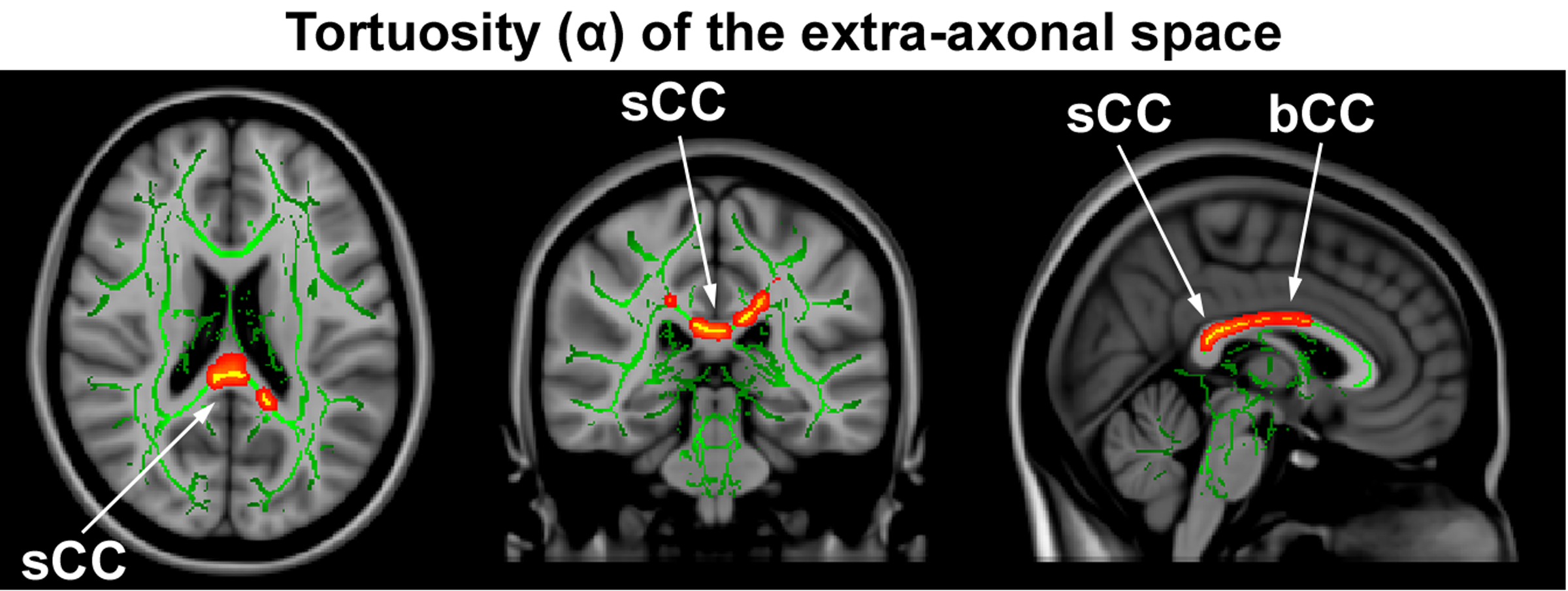
Figure 1 shows the spatial distribution of the TBSS analysis for α, particularly in the body (bCC) and splenium (sCC) of the corpus callosum (CC) (p<0.05, FWE-corrected). Other metrics did not show areas of significant difference surviving FWE correction. Based on TBSS results, ROI analysis was performed on the bCC and sCC. We observed: 1) in the bCC, significantly decreased AK and α in the MTBI (AK=0.45±0.03, 0.43±0.03 and α=2.74±0.10, 2.65±0.11); and 2) in the sCC, significantly decreased De,ǁ and α in the MTBI (De,ǁ=2.78±0.12, 2.71±0.15 and α=3.07±0.13, 2.93±0.14); NC vs MTBI, respectively. Shannon entropy was also significantly different in the bCC (AKentropy=5.24±0.07, 5.30±0.09 and αentropy=5.11±0.09, 5.01±0.14) and in the sCC (αentropy=5.08±0.09, 5.00±0.13); NC vs MTBI, respectively.DISCUSSION
We demonstrate highly localized lower α in MTBI in the posterior bCC and sCC (Fig.1). This finding is in keeping with prior results showing the CC, particularly the sCC, to be specifically susceptible to trauma, relating to shear strain forces of MTBI.5 The decreased α in MTBI is largely due to a decrease in De,ǁ (p=0.03, FWE-uncorrected), suggesting increased restrictions along the axons outside (e.g., misalignment of fibers) and/or an increase in De,﬩ (p=0.1, FWE-uncorrected), suggesting demyelination. Shannon entropy of α is also decreased in these regions, suggesting a loss of the normal heterogeneity of α seen in this region.6CONCLUSION
This study shows that there are both microstructural changes as well as regional textural changes after MTBI affecting the bCC and sCC within 4 weeks of injury. We demonstrate the potential for modeled WMTI metrics such as α, and regional texture such as Shannon entropy to be useful as early biomarkers of MTBI-related WM injury. WMTI metrics may be more sensitive to subtle injury than traditional, empiric diffusion metrics such as FA and provide further insight into the mechanisms that underlie tissue damage.Acknowledgements
Supported in part by R01 NS039135-11, R21 NS090349 and P41 EB017183.References
1. Vanderploeg RD, et al. Long-term morbidities following self-reported mild traumatic brain injury. J Clin Exp Neuropsychol. 2007;29(6):585-598.
2. Fieremans E et al. White matter characterization with diffusional kurtosis imaging. Neuroimage. 2011;58(1):177-188.
3. Smith SM et al. Tract-based spatial statistics: voxelwise analysis of multi-subject diffusion data. Neuroimage. 2006;31(4):1487-1505.
4. Shannon CE. The mathematical theory of communication. MD Comput. 1997;14(4):306-317.
5. Rutgers DR et al. Diffusion tensor imaging characteristics of the corpus callosum in mild, moderate, and severe traumatic brain injury. AJNR. 2008;29(9):1730-1735.
6. Chang YF. Possible entropy decrease in biology and some research of biothermodynamics. NeuroQuantology. 2013;11(2):189-196.
Figures