4525
Fronto-Parietal Brain Metabolites Changes Following Traumatic Brain InjuryPing-Hong Yeh1, Chen-Haur Yeh1, Gerard Riedy1, Wei Liu1, Grant Bonavia1, and John Ollinger1
1National Intrepid Center of Excellence, United States, MD, United States
Synopsis
Changes of the fronto-parietal brain metabolites following traumatic brain injury can be reflected in cognitive performance and self-reported psychological function. These results suggest that MRSI might be sensitive to the disturbance of brain metabolites in chronic military mTBI.
Introduction
Studies using magnetic resonance spectroscopy imaging (MRSI) have shown that altered brain metabolites can be detected following traumatic brain injury (TBI). The goal of this study is to assess brain metabolite changes over the fronto-parietal region in patients with a clinical diagnosis of mild TBI (mTBI), and evaluate the association between brain metabolites, neuropsychological symptoms, and cognitive function. The goal of this study is to assess brain metabolite changes over the fronto-parietal region in mild TBI (mTBI), and evaluate their association between clinical symptoms, and cognitive function.Methods
Participants and Imaging acquisition Five hundred and thirty-four active duty service members (age: 34.9 ± 8.1 years old, M/F = 511/15, average 337.3 ± 229.9 with range of [29, 996] days since injury) who had been previously diagnosed with mTBI, received a series of neuroimaging exams at the National Intrepid Center of Excellence (NICoE) using a GE MR750 3T scanner equipped with a 32 channel head coil. Fifty-six non-TBI controls were recruited for comparison. The 2D H1 chemical shift imaging (CSI) scans were acquired using a point resolved spectroscopy (PRESS) with a field of view covering the supracallosal fronto-parietal region (Fig 1A). Image analysis Post-processing of MRSI included apodization using 6Hz Lorentzian filtering, fast Fourier transformation, and phase correction followed by spectra combination of different coils (Fig. 1B,C). Peak heights of metabolites, including N-acetyl aspartate (NAA), choline (Cho) and creatine (Cr) were quantitated. Normalized values of NAA/Cr, NAA/Cho and NAA/Cr were used for further statistical analyses. Mean metabolite peak ratios of the fronto-parietal regions of interest (ROIs), based on structural T1 brain parcellation using Freesurfer (Fig. 1D), were quantitated. Statistical analysis Linear mixed modeling was applied to evaluate the difference of mean metabolite peak ratios among groups (non-TBI vs mTBI (sub)groups, including blast mTBI and non-blast TBI), while considering covariates of age and gender. Significance was tested at an uncorrected p < 0.05 because the number of ROIs were less than 10. The relationships between fronto-parietal metabolite peak height ratios, neuropsychological symptoms, and cognitive testing were evaluated using Pearson partial correlation.Results
Compared to non-TBI controls, the mTBI group had lower NAA/Cho peak height in the region of the left pars opercularis, and lower Cho/Cr over the left frontal white matter (p < 0.05). The non-blast mTBI subgroup had decreased peak height of NAA/Cho when compared to non-TBI controls over the posterior central gyrus. In addition, the blast mTBI subgroup had higher NAA/Cho peak height in the anterior cingulate, but lower NAA/Cho in the posterior cingulate than the non-blast mTBI group. Higher Cho/Cr in the left cingulum correlated with better performance of Trail Making A test (r=0.27, p=0.04) (Fig. 3). Mean NAA/Cho peak heights of the bilateral fronto-parietal white matter correlated with shorter (quicker) hit reaction time of Conners Continuous Performance Test (r = -0.28, p < 0.003). Additionally, lower NAA/Cho peak height over the left precuneus gyrus correlated with a higher score on the Post-traumatic Stress Disorder (PTSD) Checklist –Civilian version (PCL-C), signifying greater PTSD symptoms.Discussion and conclusions
These results suggest that MRSI might be sensitive to the disturbance of brain metabolites in chronic military mTBI. The difference of NAA/Cho changes in the cingulate subregions, i.e. anterior vs posterior, between blast mTBI and non-blast mTBI indicate different mechanisms of brain insults. Changes of the fronto-parietal brain metabolites can be reflected in cognitive performance and self-reported psychological function. In conclusion, MRSI can be useful in monitoring brain recovery in chronic military mTBI patients.Acknowledgements
Disclaimer: The views expressed in this article are those of the author and do not reflect the official policy of the Department of Army/Navy/Air Force, Department of Defense, or U.S. Government.
References
No reference found.Figures
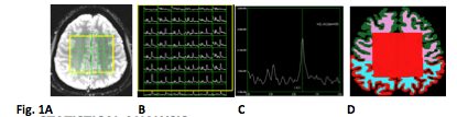
2D
H1 chemical shift imaging using
a
point resolved spectroscopy (PRESS) with a FOV covering supra-callosal fronto-parietal
region
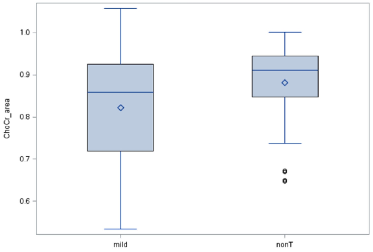
mTBI had
lower Cho/Cr than nonTBI
group over the left frontal white matter.
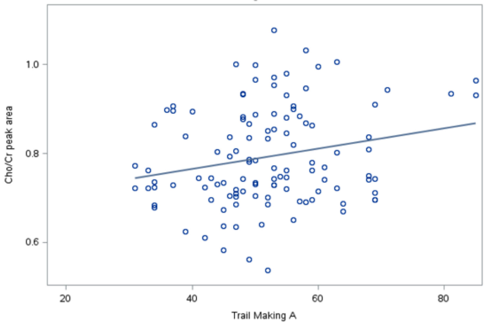
Higher
Cho/Cr in the left cingulum correlated
with better performance of Trail Making A test (r=0.27, p=0.04).