4490
Multi-phase contrast-enhanced magnetic resonance imaging with respiratory gating in papillary thyroid carcinoma1Radiology, Central Hospital of Minhang District, Fudan University, Shanghai, People's Republic of China, 2Radiology, Central Hospital of Minhang District, Fudan University, People's Republic of China
Synopsis
Purpose To investigate the role of multi-phase contrast-enhanced MR imaging in papillary thyroid carcinoma (PTC). Methods A consecutive series of 62 lesions with PTC were prospective evaluated by MR imaging. Lesions were evaluated for location, size, shape, margin, rim, degree and pattern of enhancement. Results 52 (83.9%) lesions showed moderate enhancement. 45 (72.6%) lesions showed ring-enhancement and 42 (67.7%) showed central washout enhancement during the delayed phase. 36(58.1%) manifested ill-defined margin and irregular rim (49, 79.0%) after administration of the contrast medium. Nine nodules (14.5%) showed extrathyroidal extension. Conclusion PTC commonly manifest moderate enhancement during the initial phase, with ill-defined margin and irregular rim. Central wash out enhancement and ring-enhancement occur during delayed contrast –enhanced phase.
Introduction/ Purpose
Recently, magnetic resonance imaging (MRI) studies of thyroid nodules revealed different thyroid tumors, especially papillary thyroid carcinoma(PTC) and benign nodules. Current clinical MRI studies involving thyroid focus on DWI with ADC values . However, a few recent studies suggested that malignant thyroid nodules usually have a lower ADC value compared with benign nodules, but the results show discrepancies occasionally. Additional, poor cervical MRI images show motion artifacts. We studied the decrease in motion artifacts using the respiratory gating technique. To the best of our knowledge, few studies investigated the relationship between enhanced thyroid nodules and the underlying histopathology. The purpose of our study was to evaluate the role of multi-phase contrast-enhancement in the diagnosis of PTCs.Methods
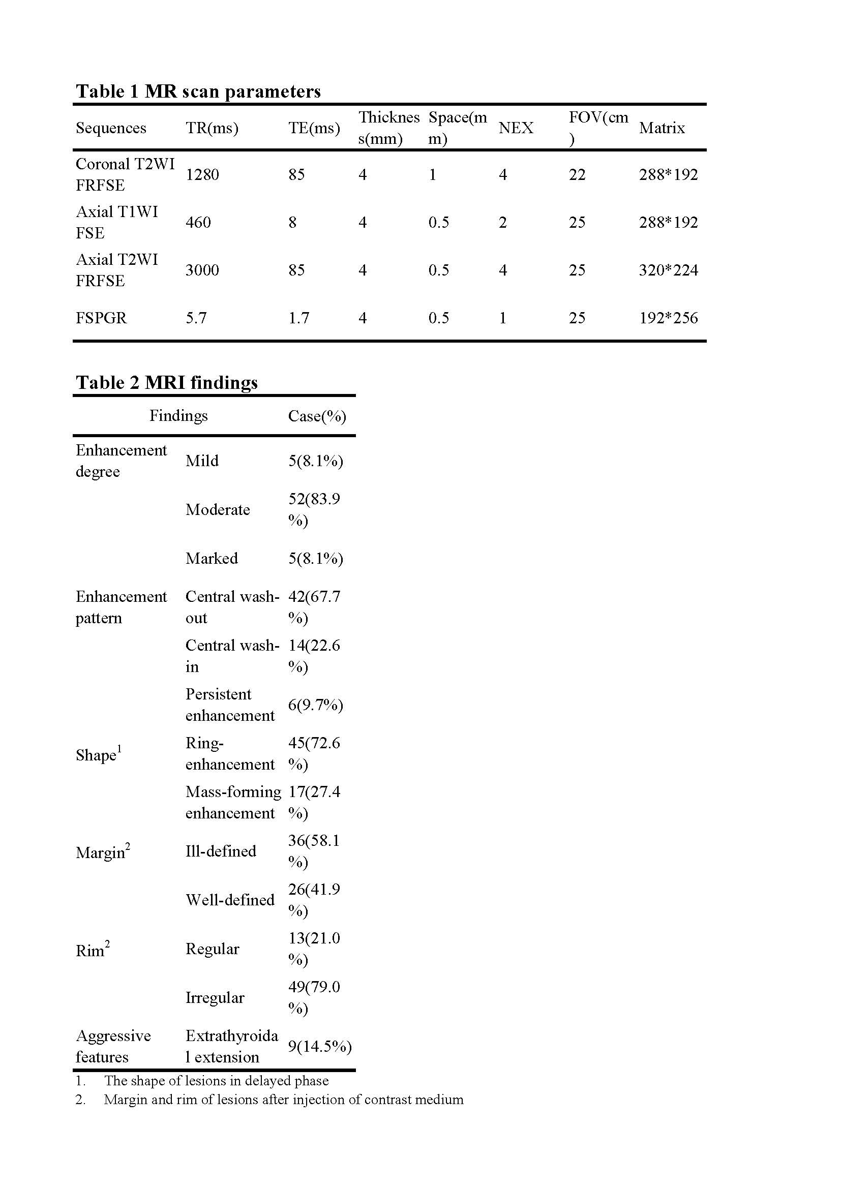
A prospective study was conducted involving 62 consecutive patients with 62 solid thyroid nodules, following multi-phase enhanced MRI of the thyroid gland. All cases were confirmed pathologically following surgery. Enhanced MR imaging was performed using a fast spoiled gradient recalled echo (FSPGR) with six phases after IV injection at the following time points: 30, 60 s, 120 s, 180 s, 240 s, and 300 s, respectively. Lesions were evaluated for location, size, shape, margin, rim, degree and pattern of enhancement.Results
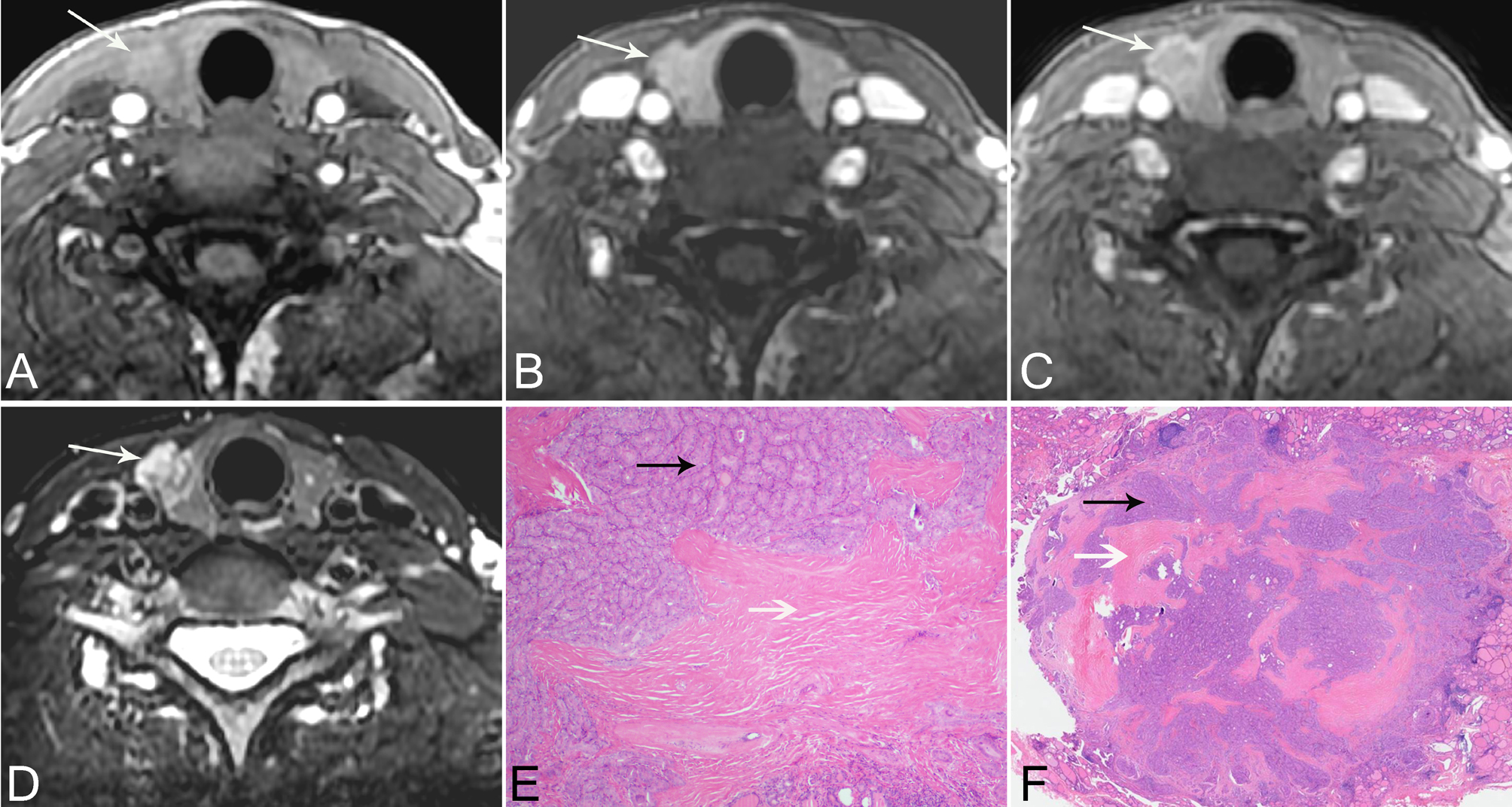
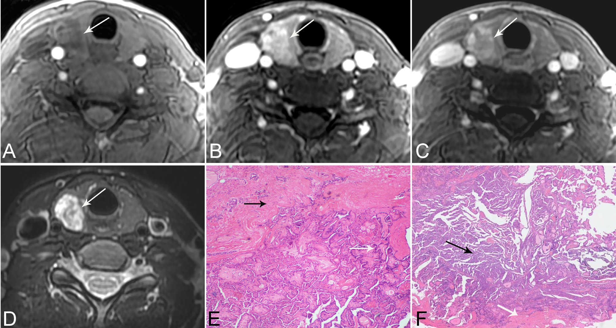
Five nodules (8.1%) showed mild enhancement, and 52 (83.9%) nodules showed moderate enhancement. Five nodules (8.1%) showed marked enhancement during the initial phase; 45 (72.6%) nodules showed ring-enhancement and the remaining 17 (27.4%) revealed mass enhancement during the delayed phase. During the delayed phase, central washout enhancement was observed in 42 (67.7%) nodules, central wash-in was seen in 14 (22.6%) nodules and the remaining six (9.7%) nodules manifested persistently enhanced pattern. Most nodules (36, 58.1%) manifested ill-defined margin and irregular rim (49, 79.0%) after administration of the contrast medium. Nine nodules (14.5%) showed extrathyroidal extension.Discussion
To the best of our knowledge, no studies evaluated the relationship between MRI enhancement pattern of thyroid nodules and the underlying histopathology. Different mechanisms were proposed to explain the varying morphology of PTCs. In our study, 52(83.9%) nodules showed moderate enhancement and five (8.1%) revealed marked enhancement during the initial phase. Increased cell density, severe desmoplastic response, neovascularity, and cell proliferation in PTCs contributed to moderate or marked enhancement. During the delayed phase, a central washout enhancement was seen in 42(67.7%) nodules, and ring enhancement in 45(72.6%) nodules during the delayed phase. The tumor area with washout indicates active growth of tumor cells, whereas the peripheral area is composed mainly of loose connective tissue with abundant intercellular matrix. Peripherally enhanced areas in PTCs during the delayed phase may also be related to fibrous stroma of the tumor and the presence of vascular fibrotic stroma. Five PTCs showed central wash-in enhancement. Fibrous scar was always seen in the central area of lesions and abundant tumor cells infiltrating peripheral area. The degree of tumor enhancement in the delayed phase image, which is usually obtained 3 to 5 min after injection of contrast medium, is closely related to the degree of interstitial space in the fibrous stroma. Some nodules (36, 58.1%) manifest ill-defined margin, irregular rim (49, 79.0%) and extrathyroidal extension (9, 14.5%) after contrast medium administration, which possibly indicate an infiltrative pattern of PTCsConclusion
Multi-phase contrast-enhanced MRI using respiratory gating improves the diagnostic imaging of PTC. PTC commonly manifests moderate enhancement during the initial phase, and ill-defined margins, irregular rim, and central wash-out and ring-enhancement in delayed contrast –enhanced phase.Acknowledgements
We thank the financial support from the Minhang district of Shanghai Science and Technology Committee (grant 2015MHZ026, 2013MHZ016).
References
1.Cappola AR, Mandel SJ. Improving the long-term management of benign thyroid nodules. Jama 2015; 313:903-904
2.Shi R, Yao Q, Wu L, et al. T2 * mapping at 3.0T MRI for differentiation of papillary thyroid carcinoma from benign thyroid nodules. Journal of magnetic resonance imaging : JMRI 2016; 43:956-961
3.Noda Y, Kanematsu M, Goshima S, et al. MRI of the thyroid for differential diagnosis of benign thyroid nodules and papillary carcinomas. AJR American journal of roentgenology 2015; 204:W332-335
4.Sasaki M, Sumi M, Kaneko K, Ishimaru K, Takahashi H, Nakamura T. Multiparametric MR imaging for differentiating between benign and malignant thyroid nodules: initial experience in 23 patients. Journal of magnetic resonance imaging : JMRI 2013; 38:64-71
5.Morris LG, Sikora AG, Tosteson TD, Davies L. The increasing incidence of thyroid cancer: the influence of access to care. Thyroid : official journal of the American Thyroid Association 2013; 23:885-891
6.American Thyroid Association Guidelines Taskforce on Thyroid N, Differentiated Thyroid C, Cooper DS, et al. Revised American Thyroid Association management guidelines for patients with thyroid nodules and differentiated thyroid cancer. Thyroid : official journal of the American Thyroid Association 2009; 19:1167-1214
Figures