4484
In-vivo targeting and imaging of super-paramagnetic iron-oxide particles to subcutaneous tumour modelsMohammad Mohseni1, John Connell1, Stephen Patrick1, May Zaw-Thin1, Tammy Kalber1, Tom Roberts1, Quentin Pankhurst2, Mark Lythgoe1, and Bernard Siow1
1CABI, UCL, london, United Kingdom, 2UCL, london, United Kingdom
Synopsis
Magnetic targeting of drug-conjugated iron oxide nanoparticles has the potential to increase the concentration of therapeutic agents to tumours whilst reducing off-target side effects of current chemotherapy methods. This preclinical work demonstrates that SPION accumulation can be increased in subcutaneous tumours using magnetic fields and can then be detected by MRI. In addition, key physiological parameters can be measured before magnetic targeting for future optimisation of the strategy.
Introduction
One of the current challenges in clinical chemotherapy is delivering a high concentration of drug to the tumour tissue whilst reducing systemic dosing and limiting off-target side effects. Magnetic nanoparticles, in combination with applied magnetic fields, can non-invasively guide small molecule drugs1, antibodies and cells2 to a specific site within the body to enhance their drug delivery efficacy. Importantly, magnetic nanoparticles also act as a MRI contrast agent, enabling non-invasive imaging of particle delivery to the region of interest. Currently, limited work has been carried out targeting nanoparticles to tumours after intravenous administration, or how physiological parameters, such as blood perfusion and vessel permeability effects. In this study we combine imaging with magnetic targeting to assess delivery of magnetic nanoparticles as a therapeutic approach to cancer therapy.Methods
In this proof of principle study, 1.5 million SW1222T human colorectal tumour cells were subcutaneously injected into nude CD-1 mice (n=6) on each flank. After a period of 14 days, super-paramagnetic iron-oxide nanoparticles, Fluid Mag-CT particles (Chemicell, 100nm diameter with a magnetite core with carboxyl functional group) were intravenously injected into the mouse and a 1T neodymium magnet was positioned over the surface of one of the bilateral tumours for 20 minutes. T2-weighted and T2* map MR images were acquired before and after particle administration using respiratory-triggered fast spin echo and multiple gradient echo sequences respectively. In addition, dynamic contrast enhanced MR images (DCE, 2D gradient echo sequence with 1 second temporal resolution) and perfusion maps (flow sensitive alternating inversion recovery with gradient echo with Look-Locker readout3 were acquired to assess blood vessel permeability and tumour perfusion using a 9.4T MRI scanner (Agilent Technologies Inc, USA). Data were analysed with in-house MATLAB code.Results
We observed notable hypointensities in all tumors with magnetic targeting due to the accumulation of iron oxide nanoparticles. As expected, these regions tended to be more prevalent when close to the magnet. Prussian blue staining on histology confirmed this. We also observed a reduction in T2* of magnetic targeting (p<0.05 t-test;n=6) compared with control (no magnet) tumour (Fig1). Quantitative perfusion mapping of subcutaneous tumours demonstrated a low and heterogeneous perfusion (0.8 +/- 1.8 ml/g/min), and gadolinium enhanced-DCE indicated a slow wash in rate (0.175 s-1) as opposed to the higher wash in rate of muscle tissue (6.57 s-1).Discussion
The results presented here show that relatively small (100nm) SPIONs accumulate in subcutaneous tumours when exposed to a magnetic field (Figure 1). This indicates that nanoparticles can be selectively steered out of vascular bed into the tumour to provide a mechanism to deliver a therapeutic payload. In the future, quantitative MRI methods for perfusion mapping and gadolinium-enhanced MRI will be used to evaluate physiological parameters, perfusion and blood vessel permeability, to tailor the magnetic targeting to the tumour.Conclusion
Static magnetic fields can increase SPION localisation in tumours. These data support the use of targeted magnetic nanoparticles using the gradient field of MRI scanner in a technique called Magnetic Resonance Targeting (MRT)4. In addition, gadolinium-enhanced MRI may be used to predict and optimise the MRT parameters.Acknowledgements
No acknowledgement found.References
(1) Vicky V. Mody Arthur Cox Samit Shah et al., Magnetic nanoparticle drug delivery systems for targeting tumor, Applied Nanoscience, 2014; 385–392 (2) Riegler J, Lau KD, Garcia-Prieto et al., Magnetic cell delivery for peripheral arterial disease: A theoretical framework, Med Phys, 2011; 38, 3932 (3) Ramasawmy, R., Campbell-Washburn, A.E., Wells, J.A et al., Hepatic arterial spin labelling MRI: an initial evaluation in mice, NMR Biomed . 2015; 28(2):272-280 (4) Munitta Muthana, Aneurin J. Kennerley, Russell Hughes et al., Directing cell therapy to anatomic target sites in vivo with magnetic resonance targeting, Nature Comms. 2015; 10.1038Figures
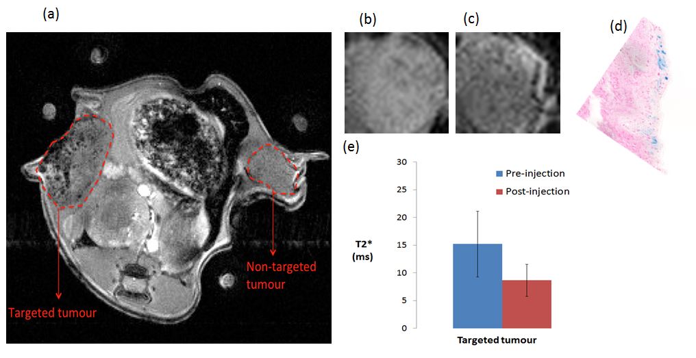
a) T2* weighted image of the mouse after particle administration
and targeting. The darker area in the targeted tumour as opposed to the
non-targeted tumour confirms the retention of particles in the targeted tumour.
(b & c) T2* weighted images of a subcutaneous tumour (b) before and
(c) after magnetic targeting. (d) Prussian blue histological staining of the
same tumour confirms particle accumulation, which is more prevalent closer to
the magnet. (e) The mean T2*value across the targeted tumour is significantly
decreased between the pre and post acquisitions (P = 0.0313 paired two-tailed Wilcoxon
test; mean±SD).