4461
Voxel-wise meta-analysis of resting-state functional activity in type 2 diabetes1Henan Provincial People’s Hospital, Zhengzhou, People's Republic of China, 2Huazhong University of Science and Technology, Wuhan, People's Republic of China, 3Department of Radiology, West China Hospital of Sichuan University, Zhengzhou, People's Republic of China
Synopsis
Previous studies about resting-state functional activity reported that altered brain activity was demonstrated with type 2 diabetes and their findings were inconsistent ,which have not been quantitatively reviewed. Therefore, we conducted a quantitative meta-analysis including resting-state functional activity studies of patients with type 2 diabetes. Meta-analysis of pooled and subgroup meta-analyses found that type 2 diabetes patients showed abnormal brain activity mainly in default mode network(DMN) and visual-related regions. Our finding supports that type 2 diabetes could lead to diabetic brain activity alterations, which may correlate with cognitive impairment of type 2 diabetes patients.
Target audience: Researchers interested in functional MRI.
Purpose: The aim of this study was to conduct a quantitative meta-analysis of studies that included the amplitude of low-frequency fluctuation (ALFF) and regional homogeneity (ReHo) in patients with type 2 diabetes.
Introduction: Recent longitudinal studies have shown that individuals with insulin resistance have an associated increased risk of developing Alzheimer disease (AD) compared to healthy individuals [1]. However, the mechanism by which the insulin resistance of type 2 diabetes patients influences brain function through acting on specific brain regions has not been fully elucidated. Previous studies on resting-state functional activity have found that patients with type 2 diabetes demonstrate altered brain activity, but these findings are inconsistent and have not been quantitatively reviewed.
Methods: We selected relevant studies published before July 2016 in PubMed, Web of Knowledge, and Ovid. Six ALFF data sets and 5 ReHo data sets were included. The seed-based mapping (SDM) method was applied to quantitatively estimate regional neural activity abnormalities in type 2 diabetes patients. Seed-based mapping is a relatively new voxel-based meta-analytic tool that has been shown to be better than previous coordinate-based meta-analytical methods, such as the activation likelihood estimation (ALE) [2]. A systematic voxel-based jackknife sensitivity analysis at the whole-brain level was conducted to test the replicability of the results. And SDM has not been used in a meta-analysis of studies exploring functional activity alterations in patients with type 2 diabetes compared to healthy controls. Type 2 diabetes was diagnosed according to the latest criteria of diabetes in all included studies.
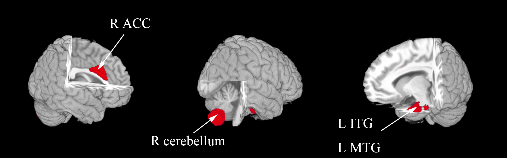
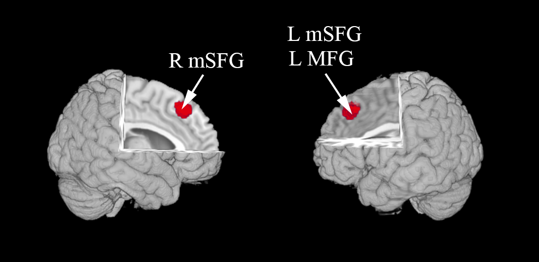
Results: A meta-analysis of 6 ALFF data sets (including 136 patients and 142 healthy controls) and 5 ReHo datasets (including 116 patients and 98 healthy controls) were conducted. Our study found that ALFF was higher in the left inferior temporal gyrus (ITG) and middle temporal gyrus (MTG) , right anterior cingulate cortex(ACC) and cerebellum of patients with type 2 diabetes compared to healthy controls (Figure 1), whereas ReHo was higher in the bilateral medial superior frontal gyrus(mSFG) and left middle frontal gyrus (MFG) (Figure 2) .
Discussion: The pooled meta-analyses revealed higher ALFF in the left MTG and ITG, right ACC and cerebellum in patients with type 2 diabetes relative to that in controls. The MTG, ITG and ACC all belong to the default mode network (DMN), which contributes significantly to normal cognitive function [3]. In addition to the regions within the DMN, increasing evidence shows that the cerebellum not only plays a pivotal role in motor functions but is also involved in cognitive, emotional, and sensory processing[4, 5]. On the other hand, the pooled meta-analyses revealed higher ReHo in the bilateral mSFG and left MFG of type 2 diabetes patients compared to that of healthy controls, which belong to the default mode network(DMN) or anatomically connected to DMN area. The altered ReHo and ALFF in these brain regions may be related to impairments in cognitive and visual function. However, this present meta-analysis study had limitations. First, there were only a small number of studies that met our inclusion criteria, which may reduce the ability to detect more neural activity changes. Second, the accuracy of our voxel-wise meta-analyses may be limited, as they should be based on raw statistical brain maps instead of the coordinates from published studies; however, all of the voxel-wise meta-analyses were performed previously as described here.
Conclusion: Despite the limitations mentioned above, our voxel-wise meta-analyses found higher resting-state ALFF in the left ITG, left MTG, right ACC and right cerebellum and higher ReHo in the bilateral mSFG and left MFG in patients with type 2 diabetes compared with healthy controls using the SDM method. The altered ReHo and ALFF in these brain regions may be related to impairments in cognitive and visual function. Furthermore, the subgroup meta-analyses suggest that SVD and type 2 diabetes have a complex interaction in the decline of cognitive and visual functions, which still needs to be explored further in a large sample with a more accurate diagnosis of the stage of type 2 diabetes.
Acknowledgements
This study was funded by National Natural Science Foundation of China (No. 31470047, 81271565).
References
1. Schrijvers, E.M., et al., Insulin metabolism and the risk of Alzheimer disease: the Rotterdam Study. Neurology, 2010. 75(22): p. 1982-7.
2. Radua, J., et al., A new meta-analytic method for neuroimaging studies that combines reported peak coordinates and statistical parametric maps. Eur Psychiatry, 2012. 27(8): p. 605-11.
3. Buckner, R.L., J.R. Andrews-Hanna, and D.L. Schacter, The brain's default network: anatomy, function, and relevance to disease. Ann N Y Acad Sci, 2008: p. 011.
4. Schmahmann, J.D., J.B. Weilburg, and J.C. Sherman, The neuropsychiatry of the cerebellum - insights from the clinic. Cerebellum, 2007. 6(3): p. 254-67.
5. Stoodley, C.J. and J.D. Schmahmann, Functional topography in the human cerebellum: a meta-analysis of neuroimaging studies. Neuroimage, 2009. 44(2): p. 489-501.
Figures