4442
Virtual Reality to read MR Angiography1Clinical Science, Philips Healthcare, Beijing, People's Republic of China, 2Digital Rubic, 3Department of Radiology, Beijing Hospital
Synopsis
A first attempt in utilizing the state of art visualizing technology - Virtual Reality to read MR angiography images.
Introduction
MRI TOF (Time-Of-Flight) imaging provides an “MR angiogram” (MRA) by minimizing signal from stationary background tissues and maximizing signal from flowing blood. It can be performed by both 2D and 3D techniques. 3D TOF MRA achieves thinner, contiguous imaging sections and much higher spatial resolution than does 2D TOF MRA. It is a widely used non-invasive method that provide safer and less expensive way to assess the stenosis or malformation of intracranial arteries(1, 2),carotid arteries(3), renal artery(4) and peripheral arteries(5) . MRA is a useful screening tests for exclusion of arterial stenosis, but is unreliable to establish the diagnosis and estimate the severity of stenosis(1). 3D-TOF is also suitable for veins and named MRV (Magnetic Resonance Venography). Contrast-enhanced or postcontrast TOF MRA use the contrast agents to decrease the saturation effects and improve the delineation of slow flow , and is able to correctly identify stenosis or occlusion (6).
MRA images can be processed by Maximum Intensity Projection (MIP) to interactively create different projections along preferred directions for 3D visualization. MIP connects the high intensity dots of the blood vessels in three dimensions from of 3D structure to 2D plane. This process can generate multiple images at different projections angles, then be displayed in a CINE format or filmed in rotation to indirectly capture the 3D depth information.
Recent surging technology Virtual Reality (VR) provides a true 3D display and can enhance image reading experience by direct 3D perception and interactive operations. Radiologist as well as patients can perceive depth of structure in the virtual word with VR technology directly. Herein we pilot in the attempt of using VR for MRA reading, which potentially would be next trend in industry and applications.
Methods
TOF MRA images, from a patient with tumor, were collected on a 3.0T MR Scanner (Ingenia, Philips Healthcare, Best, the Netherlands) by an radiologist. The raw MRA images, without MIP reformatting, were filtered to reduce noise, then a thresholding is applied then to catch vascular structures with suppressing signal from other parts of brain. Volume rendering is adapted to generate the 3D model for further polishing specifically designed for virtual reality visualization.
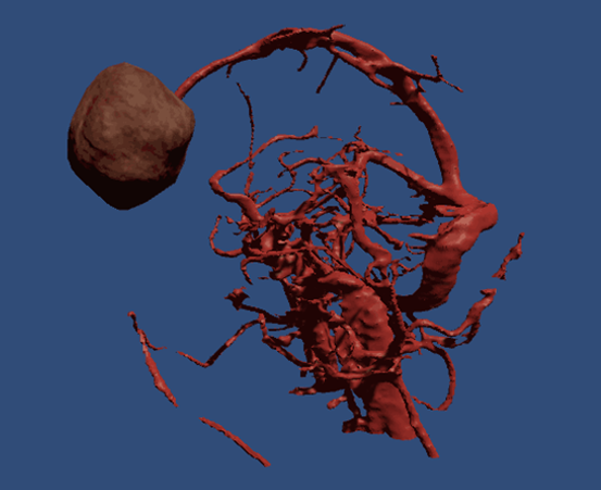
The 3D model is then refined by defining which vascular parts to be kept and volumetric noises to be eliminated. About 10^5 triangles were preserved to build up the surface of blood vessels. The surface of 3D model is then further prepared by smoothing sharp edges, followed by converting into STL format for HTC rendering engines, as shown in Figure 1.
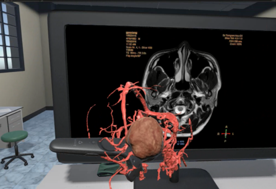
This 3D model is ready for virtual reality viewing, and which can be moved, turned as well as zoomed. However, we can’t interactively display that feature in the frame of an abstract. We also designed a virtual reading room where both tomography images can be displayed slice by slice along with the 3D blood vessel structure, to fully harvest the convince of VR environment (Figure 2). The virtual reading room is built to be capable of moving slices on the virtual monitor by the radiologist to check and compare tomography images with 3D models.
Results and discussion
An advanced image reading method is developed for angiography, and potentially could be appplied in neurography as well. With the quick and precise locating lesions and anatomy, this technology might lead a new trend in the radiology. Other potential benefits, including surgical planning and simulations, are under explorations.Acknowledgements
No acknowledgement found.References
1. Holmstedt CA, Turan TN, Chimowitz MI. Atherosclerotic intracranial arterial stenosis: risk factors, diagnosis, and treatment. The Lancet Neurology. 2013;12(11):1106-14.
2. Josephson CB, White Pm Fau - Krishan A, Krishan A Fau - Al-Shahi Salman R, Al-Shahi Salman R. Computed tomography angiography or magnetic resonance angiography for detection of intracranial vascular malformations in patients with intracerebral haemorrhage. (1469-493X (Electronic)).
3. Weber J, Veith P Fau - Jung B, Jung B Fau - Ihorst G, et al. MR angiography at 3 Tesla to assess proximal internal carotid artery stenoses: contrast-enhanced or 3D time-of-flight MR angiography? (1869-1447 (Electronic)).
4. Tan KT, van Beek Ej Fau - Brown PWG, Brown Pw Fau - van Delden OM, van Delden Om Fau - Tijssen J, Tijssen J Fau - Ramsay LE, Ramsay LE. Magnetic resonance angiography for the diagnosis of renal artery stenosis: a meta-analysis. (0009-9260 (Print)).
5. Meaney JF. Magnetic resonance angiography of the peripheral arteries: current status. (0938-7994 (Print)).
6. Ishimaru H, Ochi M Fau - Morikawa M, Morikawa M Fau - Takahata H, et al. Accuracy of pre- and postcontrast 3D time-of-flight MR angiography in patients with acute ischemic stroke: correlation with catheter angiography. (0195-6108 (Print)).
Figures