4433
Progress in adapting SWIFT to a clinical scanner1University of Minnesota, Minneapolis, MN, United States
Synopsis
The development of a programmable high-speed triggering to enable progress in implementation of SWIFT on a Siemens scanner. First in-vivo images, at 50khz, using a linear T/R coil.
Introduction
SWIFT is a silent imaging technique that has unique imaging capabilities to short T2* imaging, but has hardware requirements that exceed the capabilities of typical clinical scanners. Previous attempts to implement SWIFT on a Siemens clinical scanner were achieved, although with an extremely limited acquisition bandwidth of only 3.6 kHz [1] Implementation of SWIFT.; Here, we implemented SWIFT imaging on a Siemens 7T scanner, obtaining a more useable bandwidth of 50 kHz .Methods
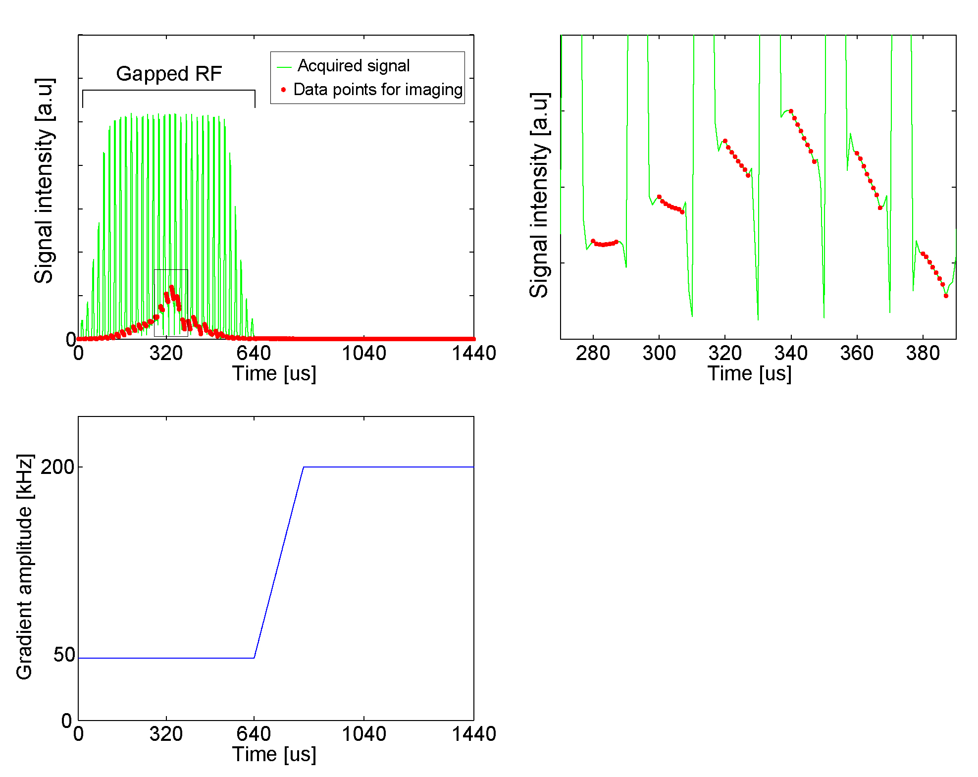
The SWIFT sequence was implemented on a standard Siemens MAGNETOM 7T system, running software version VB17A, equipped with a single-channel 8 kW amplifier for RF transmit (CPC). The sequence has a dead time of 1.5 µs before RF transmission to allow for T/R switching and RF amplifier unblanking, and an adjustable RF segment duration, chosen as 5 µs for this study. RF gapping was applied at 50 kHz, using a stretched hyperbolic secant (HS2) pulse excitation with bandwidth = 49 kHz, 32 segments and 640 µs total pulse duration. After the RF pulse, gradients are ramped to 200 kHz with a gradient rise time of 180 µs. Data was processed as in [2].
The gapped RF pulse waveforms were generated online within the pulse sequence and digitized with 1 µs resolution. Since the manufacturer's standard RF infrastructure does not support rapidly gating acquisition during a gapped RF pulse, a custom programmable high-speed triggering device was developed and integrated into the RF system of the scanner to control a fast external T/R switch (via an optical signal) and to rapidly unblank the RF power amplifier. This external triggering device is capable of generating gating control signals for the T/R switch and RF amplifier of arbitrary duration with 100 ns resolution, digitally controlled by the running pulse sequence. Data acquisition was performed without interruption throughout transmission of the gapped RF pulse and continuing afterwards for the collection of the FID signal, sampling at 1 MHz.
To allow for the required pre-amplifier protection we installed a T/R switch module (Agilent, Santa Clara, CA) with -40 dB isolation and capability to switch within 1 µs. This T/R switch was physically located at the service end of the magnet. The driver for the T/R switch module was mounted in close vicinity and equipped with a fiber optic controlled trigger input.
Results/Discussion
The rapid blanking and unblanking of the RF amplifier was evaluated and compared with having the RF amplifier unblanked during the entire duration of the gapped SWIFT pulse. The latter was found to allow significant noise into the receivers. Using the rapid blanking/unblanking of the RF amplifier combined with the fast T/R switch, data acquisition was successfully performed. In-vivo data acquisition was performed on a human subject in accordance with an IRB protocol with the University of Minnesota, using a linear transmit/receiver coil. In figure 1 (upper left), the measured signal intensity for a single FID is shown along with the data-points used for image reconstruction. A zoomed view is display on figure 1(upper right), with the red dots showing the exact datapoints used. The applied gradient readout is shown in figure 1, lower left.
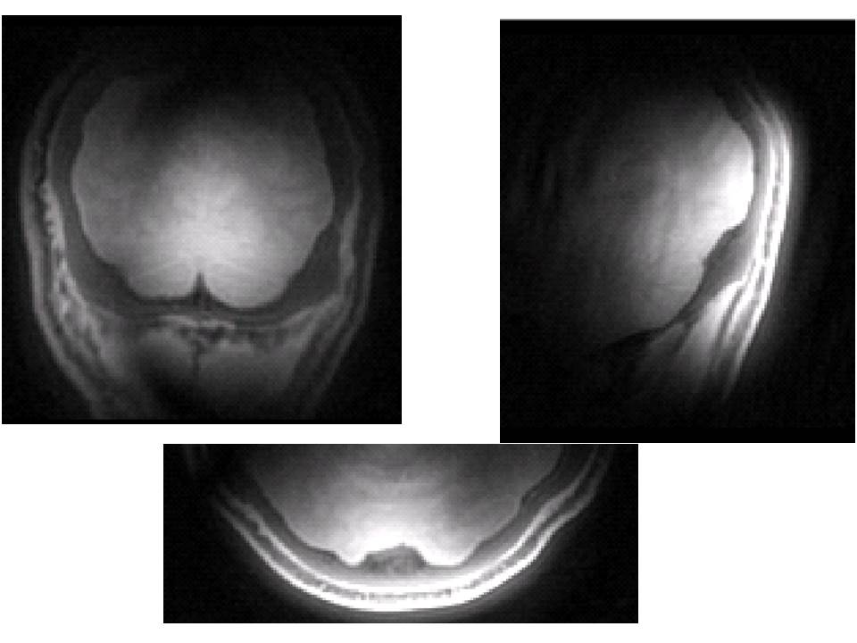
In Figure 2, axial, sagittal and coronal images of human brain are shown from an imaging experiment, exhibiting mostly PD contrast due the limited power used.The individual T/R switch module used for this study is rated for up to 500 W maximum RF power. We plan to employ up to 16 such modules in conjunction with 16-channel transmit/receive coils for future head and body work.
Acknowledgements
P41 EB015894; S10 RR026783 “Multichannel Transmit Frontend for 7 Tesla” WM KECK FoundationReferences
[1] Valette J, Moeller S, Idiyatullin D, Corum C, Le Bihan D, Garwood M, et al., ISMRM 17th Annual Scientific Meeting & Exhibition; 2009 18 -24 April; Honolulu, Hawaii USA.
[2] Kobayashi N, Lei J, Utecht L, Garwood M, Ingbar D, Bhargava M. 3D Cine Magnetic Resonance Imaging of Rat Lung ARDS using Gradient-modulated SWIFT with Retrospective Respiratory Gating. Proc SPIE Int Soc Opt Eng. 2015 Feb 21;9417. pii: 941718.
Figures