4432
An evaluation of RF coil materials for 1H/31P for use in a hybrid MR-PET scanner at 3T1Institute of Neuroscience and Medicine-4, Forschungszentrum Juelich, Juelich, Germany, 2Faculty of Medicine, Department of Neurology, JARA, RWTH Aachen University, Aachen, Germany
Synopsis
In a simultaneous MR-PET experiment, an RF resonator for MRI is placed inside the field-of-view of the PET scanner. Attenuation caused by RF coil components can cause severe artefacts in PET images if not adequately corrected. Although it may be possible to minimise subject-specific attenuation and scatter effects, correcting those due to the RF coil is still a challenging task. In this work, we have designed and constructed a number of RF coils constructed utilising a variety of copper conductors and evaluated their characteristics from the MR and PET points of view.
PURPOSE
The use of X-nuclei such as 13C or 31P with 1H MRI enables accessing valuable cellular and metabolic information. The availability of hybrid MR-PET scanners, capable of simultaneous acquisition of both datasets, has increased over the last few years. In combination with PET, X-nuclei MR has been shown to be advantageous1,2. In a simultaneous MR-PET experiment, an RF resonator for MRI is placed inside the field-of-view of the PET scanner. Standard, dual-tuned RF coils tend to be designed without considering factors such as attenuation or scatter, which are rather important effects in PET that can lead to image quality degradation. Attenuation caused by RF coil components can cause severe artefacts in PET images if not adequately corrected. Although it may be possible to minimise subject-specific attenuation and scatter effects, correcting those due to the RF coil is still a challenging task3. In this work, we built 1H and 31P RF coils using different types of copper and evaluated their characteristics from the MRI and PET perspectives.METHODS
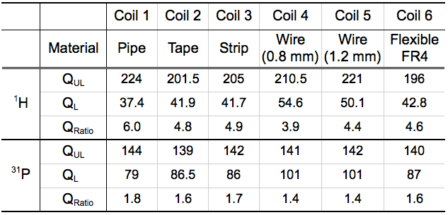
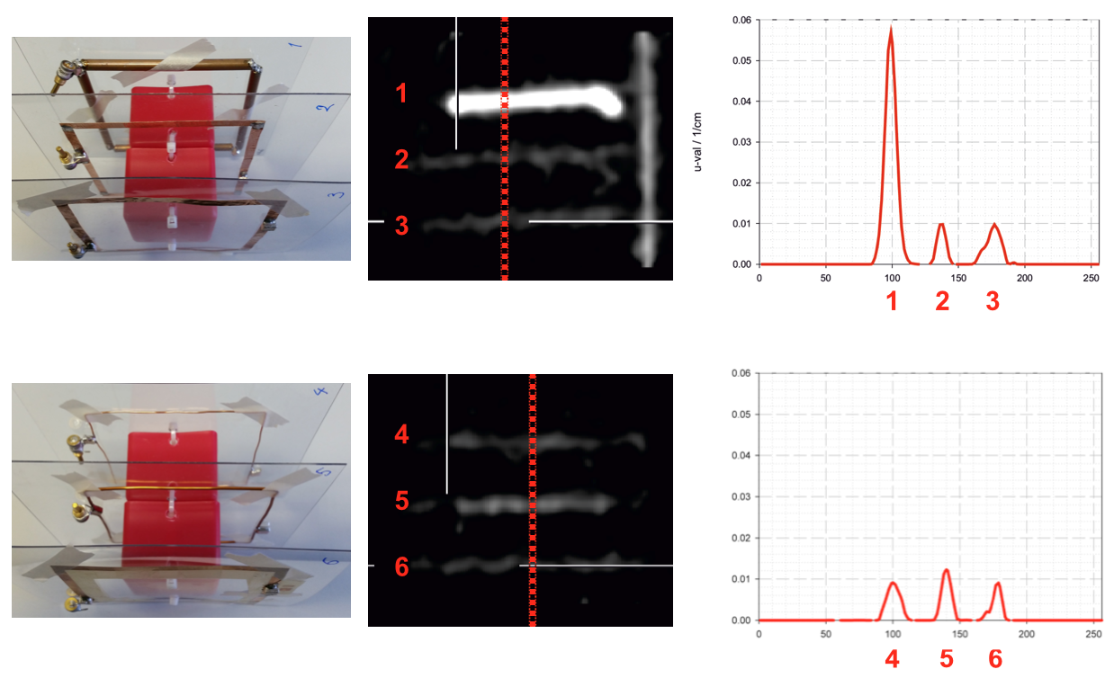
Twelve square loop coils (six each for 1H and 31P) were constructed using various types of copper – pipe, tape, strip, 0.8 mm wire, 1.2 mm wire and flexible FR4. In order to characterise their MR performance, Q-factors (loaded/unloaded/ratio) of these coils were measured on the bench using a pair of sniffer loops (45 mm diameter) placed ~28 mm away from the coils at 49.88 MHz (corresponding to 31P Larmor frequency at 3T) and 123.23 MHz (1H frequency). A uniform water phantom containing 44.8 mM NaCl and 4.7 mM NiSO4 was used for the loaded Q measurements. The PET attenuation measurements were then carried out by use of three Ge68-transmission sources (~35 MBq each) on a dedicated PET scanner (SIEMENS ECAT Exact HR+). As shown in Fig. 1, three coils were stacked with a 38 mm gap for two 30-minute transmission scans. The acquired data were reconstructed iteratively (OSEM2D, 6 iterations, 16 subsets) into 63 slices (2.45 mm) with a matrix size of 256 x 256 (1 x 1 mm) representing the value of the attenuation coefficient of the materials.RESULTS
Table 1 summaries the measured Q-factors of the coils showing that the Coil 1 built using copper pipes has the highest MR sensitivity at both 1H and 31P frequencies. The Q-factors of the Coils 2 and 3 are not as good as the Coil 1 at 1H frequency but they are comparable at the 31P frequency. On the other hand, in the transmission scan data shown in Fig. 1, the attenuation value is considerably higher (more than 5 times) for the Coil 1 while the differences of the others are negligible.DISCUSSION/CONCLUSIONS
We have evaluated the characteristics of a number of RF coils constructed utilising a variety of copper conductors from the MR and PET points of view. Since the MR sensitivity of 31P or X-nuclei is inherently low, the coil is likely to be designed with multi-layer structure4 in order to improve sensitivity. With this design, however, the selection of the type of the material may play a crucial role in finding the optimal balance that can be beneficial for both modalities5. In the future we intend to build a double-tuned coil using the copper strip optimised for both MR and PET and to conduct simultaneous 1H/31P MR-PET experiments in order to characterise it.Acknowledgements
No acknowledgement found.References
1. Shah NJ. Multimodal neuroimaging in humans at 9.4 T: a technological breakthrough towards an advanced metabolic imaging scanner. Brain Struct Funct. 2014. Jul;14.
2. Oehmigen M, Lindemann ME, Sauer M, Lanz T, Quick HH. Evaluation and integration of a dual-tuned 13C/1H headcoil into PET/MR hybrid imaging. ISMRM. 2016;3637.
3. Paulus DH, Tellmann L, Quick HH. Towards improved hardware component attenuation correction in PET/MR hybrid imaging. Phys. Med. Biol. 2013;58:8021-8040.
4. Shajan G, Mirkes C, Buckenmaier K, et al. Three-layered radio frequency coil arrangement for sodium MRI of the human brain at 9.4 Tesla. Magn. Reson. Med. 2015;75:906-916.
5. Sander CY, Keil B, et al. A 31-Channel MR brain array coil compatible with positron emission tomography. Magn. Reson. Med. 2015;73:2363-2375.
Figures