4431
Battery-Powered Integrated Parallel Reception, Excitation, and Shimming (iPRES) Head Coil Array for Plug-and-Play Localized B0 Shimming1Brain Imaging and Analysis Center, Duke University, Durham, NC, United States
Synopsis
Integrated parallel reception, excitation, and shimming (iPRES) coil arrays enable RF excitation/reception and localized B0 shimming with a single coil array, but so far use an external DC power supply and require a B0 map and a shim optimization for each subject, which takes additional time. Here, we propose a novel iPRES head coil array powered by an on-board MR-compatible battery that delivers fixed DC currents optimized in advance to shim an average subject's brain. This stand-alone, plug-and-play system does not require any subject-specific shim optimization, thus enabling a wider adoption of iPRES in clinical applications.
Introduction
Susceptibility differences at air/tissue interfaces induce static magnetic field (B0) inhomogeneities and image artifacts, which hamper many MRI applications. Conventional spherical harmonic shim coils cannot shim these localized B0 inhomogeneities, whereas multi-coil shimming1 requires an additional set of localized shim coils inside or outside the RF coil, resulting in a reduced signal-to-noise ratio or shimming performance. Integrated parallel reception, excitation, and shimming (iPRES)2 is a novel concept that allows RF and DC currents to flow in the same coil simultaneously, thereby enabling RF excitation/reception and localized B0 shimming with a single coil array, without requiring additional space in the magnet bore or compromising the signal-to-noise ratio or shimming performance.
Existing head3,4 and body5 iPRES coil arrays use a multi-channel DC power supply, which allows the currents to be optimized for each coil element, each subject, and each slice, resulting in the best shimming performance. However, this setup requires cables and filters to connect and provide RF isolation between the coil array and the power supply located outside the scanner room. In addition, its operation involves acquiring a B0 map and performing a shim optimization for each subject to determine the optimal currents, which requires additional time and may not be practical for clinical applications.
Here, we propose a novel iPRES head coil design, in which the external DC power supply is replaced by an MR-compatible battery mounted directly on the coil array, thereby eliminating cables and filters and reducing the cost and complexity of the system. The battery delivers fixed DC currents to a subset of coil elements, which are optimized in advance to shim an average subject's brain. As such, while this setup does not offer the flexibility to adjust the currents for each coil element, each subject, and each slice, it provides a stand-alone, plug-and-play system that does not require any subject-specific shim optimization, thus enabling a wider adoption of iPRES in clinical applications.
Methods
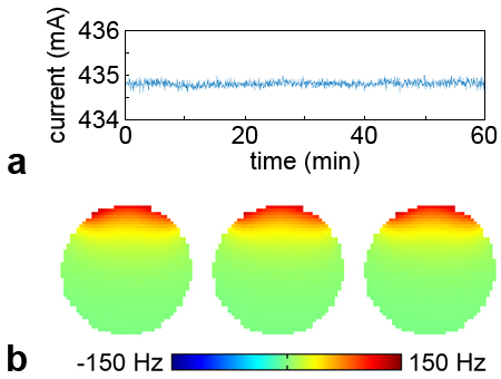
In this initial proof-of-concept implementation, an MR-compatible battery was mounted on a 32-channel iPRES head coil array3 and connected to 2 (out of 32) coil elements symmetrically located on the right and left sides of the brain. The optimal pair of coil elements and DC currents to use were determined as follows. First, basis B0 maps were acquired on a water phantom with a 1 A current separately applied in each coil element. B0 maps were also acquired in the brain of 10 healthy volunteers (7 males, 3 females, aged 18–59, with various head sizes and shapes) and were subsequently averaged. For each of the 16 pairs of right/left coil elements, the optimal current to apply was determined by minimizing the root-mean-square error (RMSE) between the average B0 map of the brain and the two corresponding basis B0 maps. The battery was then connected to the two coil elements resulting in the smallest value among the 16 RMSEs (specifically, the two inferior frontal coil elements surrounding the eyes) and was set up to deliver the optimal DC current (435 mA).
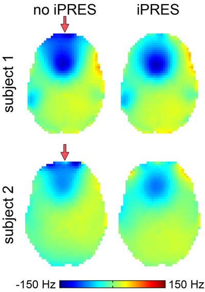
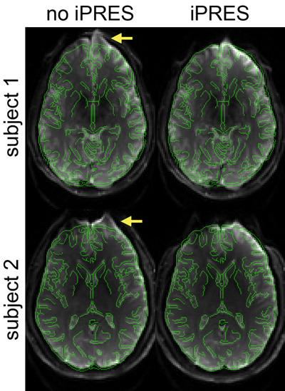
The stability of the system was tested by monitoring the current over time in bench-top measurements and by acquiring repeated B0 maps on a water phantom. The shimming performance was assessed by acquiring B0 maps and single-shot echo-planar images in two healthy volunteers, with and without DC currents applied in the battery-powered iPRES head coil array. Fast spin-echo images were also acquired for anatomical reference. All scans were performed on a 3T scanner using linear shimming, but no high-order spherical harmonic shimming.
Results and Discussion
The bench-top measurements and phantom experiments show that the DC current and B0 maps were stable (Fig. 1). Preliminary results show that shimming with the battery-powered iPRES head coil array significantly reduced the B0 inhomogeneities (Fig. 2) and geometric distortions (FIg. 3) in the frontal brain region, both for a subject who was included in the shim optimization (subject 1) and for a subject who was not (subject 2). Since DC currents were only applied in two coil elements in this proof-of-concept implementation, there were still residual B0 inhomogeneities and distortions, but the shimming performance can be further improved by powering additional coil elements with MR-compatible batteries. DIfferent settings could also be preprogrammed to shim different brain regions (e.g., frontal vs. temporal), depending on the application.Conclusion
As an alternative to using an external DC power supply and a subject-specific shim optimization, the proposed battery-powered iPRES head coil array provides a stand-alone, plug-and-play system, which will enable the adoption of iPRES in clinical applications that would otherwise not benefit from localized B0 shimming.Acknowledgements
We thank Brain Vision, LLC for their support with the MR-compatible battery. This work was in part supported by NIH grants R21 EB018951 and R24 MH106048.References
1. Juchem C et al. J Magn Reson 2011;212:280–8.
2. Han H et al. Magn Reson Med 2013;70:241–7.
3. Truong TK et al. NeuroImage 2014;103:235–240.
4. Stockmann J et al. Magn Reson Med 2016;75:441–451.
5. Darnell D et al. Magn Reson Med 2016; doi:10.1002/mrm.26267.
Figures