4428
Real-time Percutaneous Transluminal Angioplasty with Traveling Wave Magnetic Particle Imaging1Department of Diagnostic and Interventional Radiology, University Hospital Würzburg, Würzburg, Germany, 2Department of Experimental Physics 5 (Biophysics), University of Würzburg, Würzburg, Germany, 3Department of Diagnostic and Interventional Neuroradiology, University Hospital Würzburg, Würzburg, Germany
Synopsis
A percutaneous transluminal angioplasty (PTA) is a procedure to widen stenotic blood vessels in medical conditions such as coronary heart disease or peripheral artery disease. With the assistance of fluoroscopic guidance and radiopaque contrast agents, guidewires and balloon catheters are used to treat vascular stenoses. Magnetic Particle Imaging (MPI) is a very fast and sensitive tomographic imaging modality with the potential of real-time radiation-free 3D imaging. In this work the feasibility of performing a MPI-guided PTA in artificial stenoses with a traveling wave MPI scanner is demonstrated.
Purpose
Visualization of artificial stenoses in vessels phantoms, tracking of endovascular instruments and implementation of a MPI-guided PTA in real-time.Methods
Since the first publication of Magnetic Particle Imaging (MPI) in 2005 several scanner concepts have been realized 1. The traveling Wave MPI (TWMPI) approach is a scanner design, which provides a large FOV (65×25×25 mm³) 2, a rapid image acquisition (up to 2000 frames per second) 3 and a good spatial resolution (about 1.5 mm isotropic) 4. For working with MPI-guided intervention not only a fast data acquisition but also a fast image reconstruction is mandatory. In a first step the reconstruction process had to be reworked to provide a real-time preview capability of the scanned FOV. By optimization of the data transfer (data compression), the data pre-processing (using fast LAPACK, BLAS and FFTW libraries) and the reconstruction as well as the image generation step (direct deconvolution 4 and system matrix approach 5), the speed of the reconstruction software package was increased up to 10 frames per second, which is sufficient for guided interventions. To optimize the performance of the proposed method several paramagnetic marker lacquers for tracking endovascular instruments (guidewire and balloon catheter) were tested. Most suitable was a lacquer consisting of the SPIO agent Ferucarbotran embedded in a clear varnish. In the next step a vascular stenosis phantom (50 % stenosis) was constructed using a soft plastic tube with an inner diameter of 8 mm. For implementation of a short stenosis a customized ligature system was used, which provides a pressure stable ligature up to a certain pressure threshold imitating realistic conditions. Finally, the handling protocol for performing a MPI-guided PTA in an artificial stenosis phantom was optimized.Results
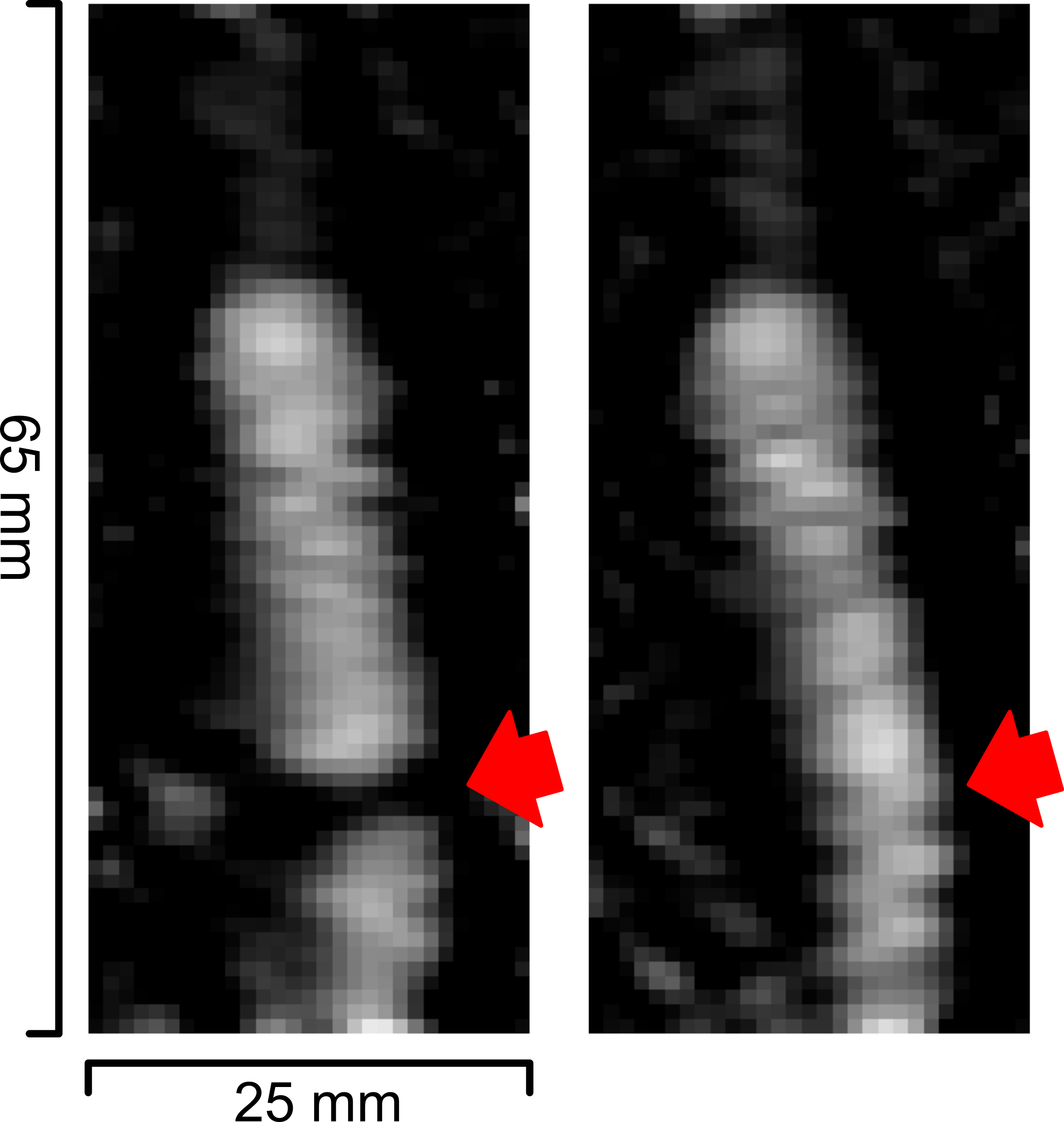
This work demonstrates the dynamic visualization of a balloon dilatation in real-time using the established MPI system. In Fig. 1 the two snapshots of the real-time visualization show the situation before and after the PTA treatment. In the left image the balloon catheter is inflated with diluted Ferucarbotran under low pressure clearly demarking the stenosis. The right image shows the widened vessel after increasing the pressure in the balloon with an insufflation pump (>10 atm).Conclusion
Magnetic particle imaging is a promising radiation-free tomographic imaging method for guided endovascular intervention, which offers imaging and visualization with a sufficient high temporal resolution to perform MPI-guided PTA.Acknowledgements
This work was partially funded by the DFG (BE-5293/1-1).References
1. N. Panagiotopoulos, et al., Magnetic Particle Imaging: current developments and future directions, Int. J. NanoMed., vol. 10, pp. 3097-3114, 2015.
2. P. Vogel, et al., Traveling Wave Magnetic Particle Imaging, IEEE TMI, col. 33(2), pp. 400-407, 2014.
3. P. Vogel, et al., Superspeed TWMPI, IEEE Trans. Magn., vol. 51(2): 6501603, 2015.
4. P. Vogel, et al., First in-vivo Traveling Wave Magnetic Particle Imaging of a beating mouse heart, Phys. Med. Biol., vo. 61(18), pp. 6620-6634, 2016.
5. P. Vogel et al., Flexible and Dynamic Patch Reconstruction for TWMPI, International Journal on MPI, vol. 2(2):1611001, 2016.
Figures