4417
A simple frequency selectable method controlled by PIN diode in the double layered multi-nuclei RF coil at 7T1Department of Health Sciences and Technology, Gachon University, Incheon, Korea, Republic of, 2Neuroscience Research Institute, Gachon University, Incheon, Korea, Republic of, 3Department of Biomedical engineering, Gachon University, Incheon, Korea, Republic of, 4Department of Radiological Science, Gachon University, Incheon, Korea, Republic of
Synopsis
With the facilitated approach of ultra-high frequency (UHF, over 7T) magnetic resonance imaging (MRI) system, multi-nuclei (MN)-MRI can be regarded as one of critical means for diagnose due to its capability of acquiring non-invasive metabolic information1, 2. In this study, a radiofrequency (RF) coil that can selectively receive two nuclei signals, the proton (1H) and the sodium (23Na), by controlling PIN diodes is proposed. Since 1H images show clear anatomical structure where 23Na show critical in-vivo metabolisms, acquired MN magnetic resonance (MR) images using the proposed MN RF coil have capabilities to assist diagnose visually3.
Purpose
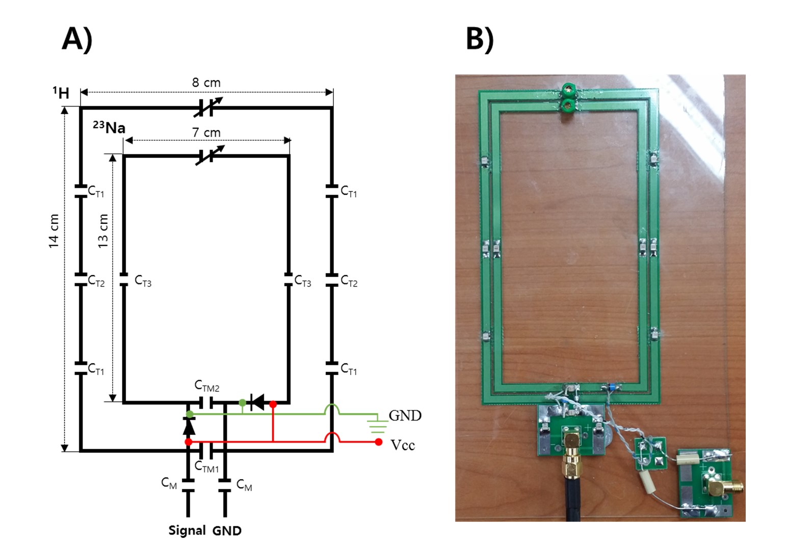
A new type of MN RF coil is proposed in this study. To acquire two different nuclei signals, ordinary method can be described as capturing 23Na(X-nuclear) signal with X-nuclear RF coil where positional information was achieved by different 1H RF coil as well as designing a single RF coil of which having two different resonance frequencies4-5. A loop type of double layered single channel RF coil having two different resonance frequencies is designed. Each layer has its own resonance frequency and can be separately driven for targeted nuclear. Proposed method can be applied to multi-channel RF coil which is capable of achieving X-nuclear signals at the specifically aimed region.Methods
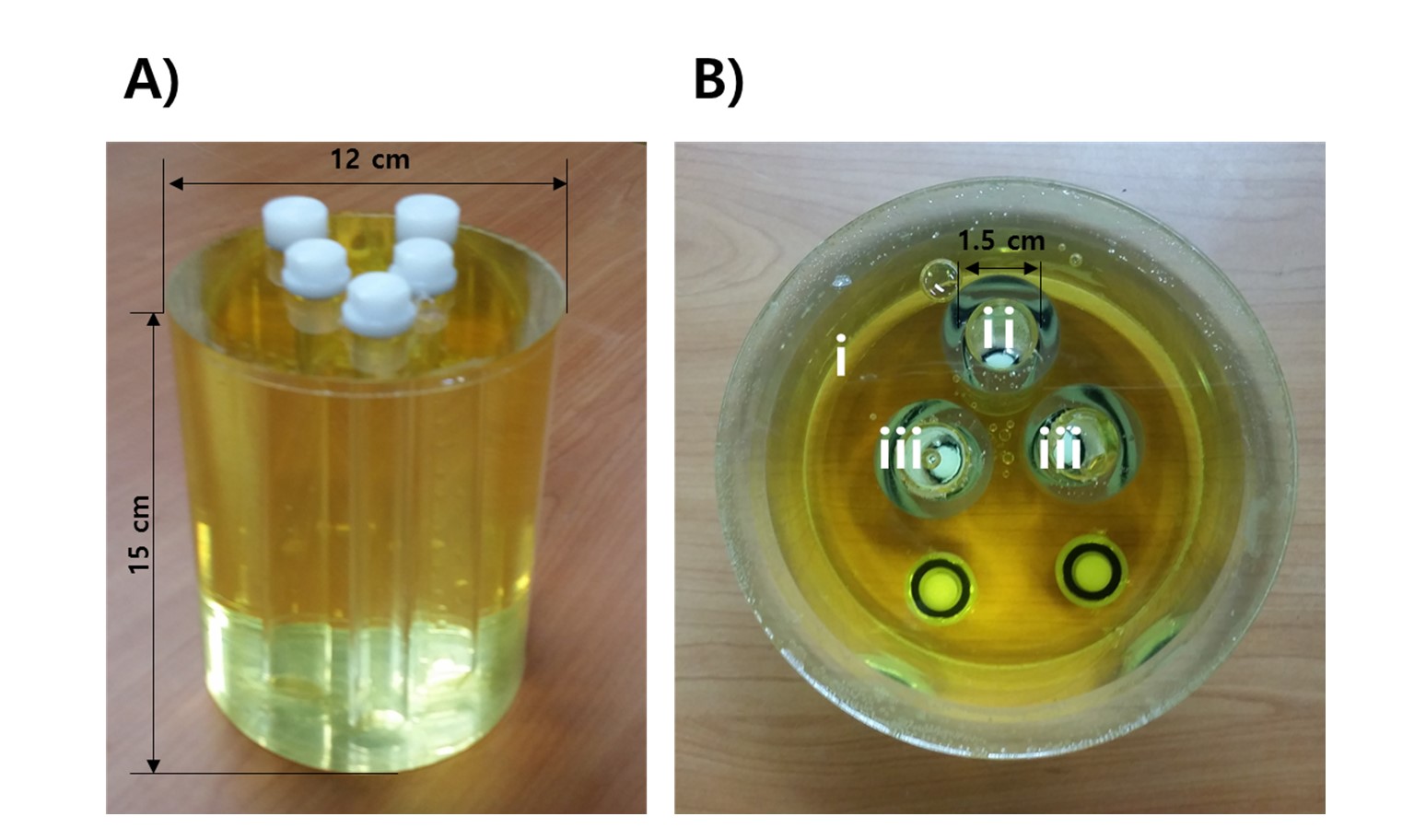
It is proposed that a certain frequency can be chosen to be received at the designed RF coil. Outer layer was tuned for 1H and inner layer for 23Na sharing the matching circuit (Fig. 1). Two PIN diodes were planned to be inserted at the source and the ground of inner layer making inner layer to be isolated without direct current (DC) supply whereas shifting resonance frequency of outer layer by voltage dip caused by the turned-on DC supply6. RF chokes were placed between the supplier and the RF coil for blocking the RF toward the DC supplier. It was proved experimentally with 7 T MR scanner (Magnetom, Siemens, Germany). A gradient echo sequence (flip angle: 90°, TR/TE: 21/11 ms, slice thickness: 8 mm and matrix size: 64 × 64) was applied to both 1H and 23Na imaging. A cylindrical phantom with three inner cylinders of triangular formation was proposed for MR imaging. The exterior cylinder was filled with oil and for the three inner cylinders, physiological saline was filled into the apex cylinder where distilled water was filled into the other two cylinders (Fig. 2).Results
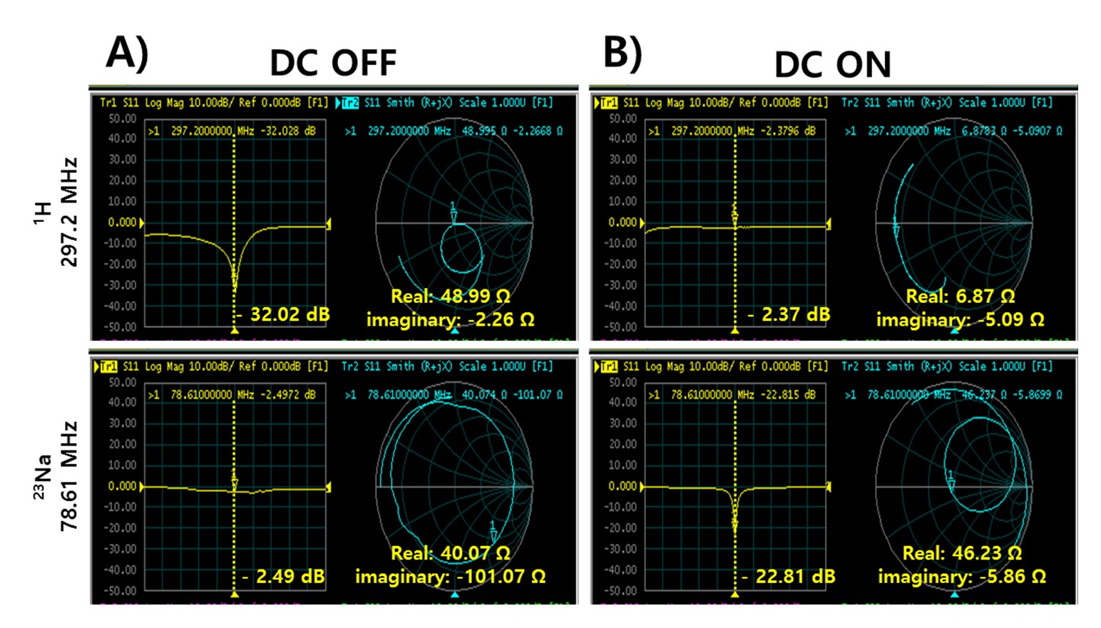
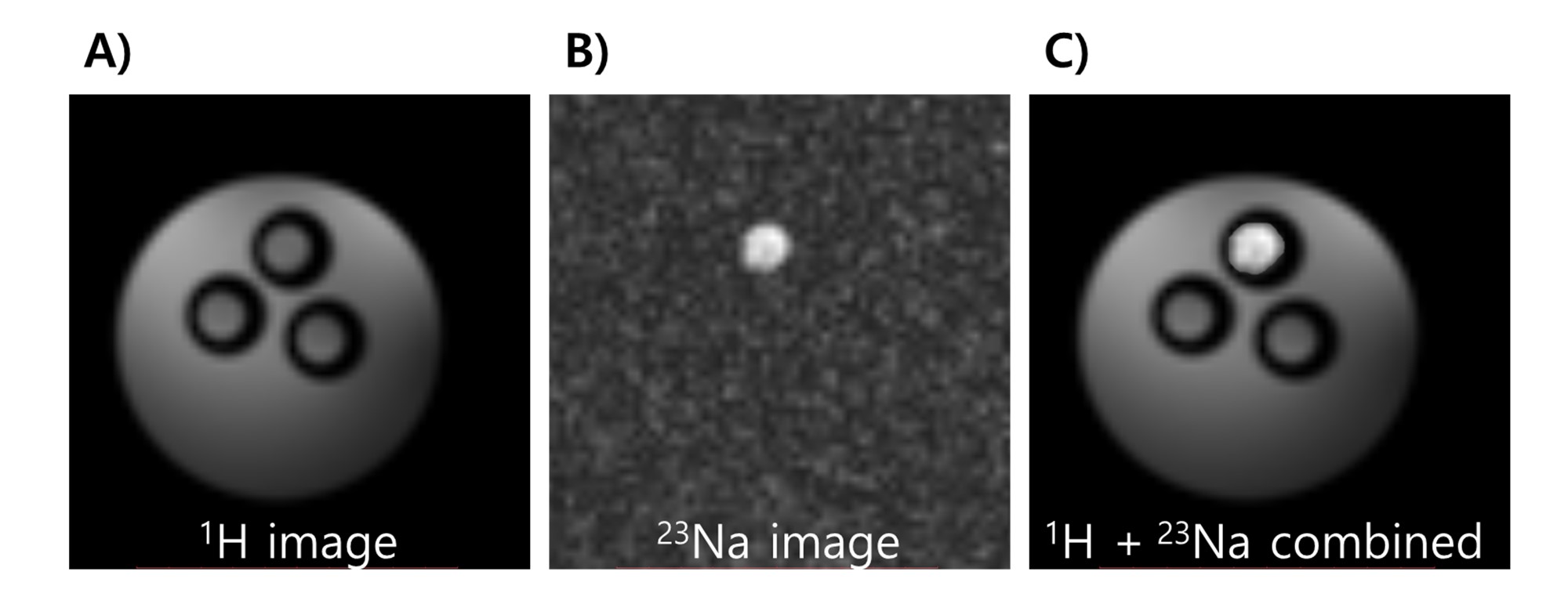
Reflection parameter (S11) profiles on a network analyzer indicate the constructed RF coil operates as proposed under the condition of DC supply control. Targeted resonance frequencies could be separated as intended (Fig. 3). With the absence of DC, S11 of 1H at the 297.2 MHz, a resonance frequency of 1H at 7 T, shows -32.08 dB where that of 23Na at the 78.61 MHz, a resonance frequency of 23Na at 7 T, shows -2.49 dB, meaning the RF coil was practically controlled to separate each resonance frequency without DC supply. With DC supply, on the contrary, S11 of 1H at the 297.2 MHz shows -2.37 dB while that of 23Na at the 78.61 MHz shows -22.81 dB. These results present that signal of only aimed frequency can be achieved by controlling DC supply. Using the previously described cylindrical phantom, experiment of proposed method was performed at the 7 T MR system. Anatomical and functional image was able to be acquired by combining 1H imaging without DC supply and 23Na imaging with DC supply confirming proposed method can be applied (Fig. 4).Discussion and Conclusion
With the proposed RF coil design, X-nuclear signals can be obtained under certain circumstances by providing DC whereas ordinary anatomical MR images also can be achieved without DC supply. There exists, however, some shortcomings such as high realization difficulty resulting from two individual layers sharing one matching circuit and necessity of DC supplier at the MR system. Despite that this study was proceeded with only single channel RF coil, it can be predicted that higher signal-to-noise ratio (SNR) and faster imaging with multi-channel RF coil making further study on the multi-channel RF coil and appropriate decoupling method must be the next objectives.Acknowledgements
This work was supported by a National Research Foundation of Korea (NRF) grant funded by the Ministry of Education, Science and Technology of the Republic of Korea (grant number: NRF-2014M3C7033998), and a grant from the Korea Health Technology R&D Project through the Korea Health Industry Development Institute (KHIDI), funded by the Ministry of Health and Welfare of the Republic of Korea (grant number: HI14C1135).References
1. Roemer PB, Edelstein WA, Hayes CE, Souza SP, Mueller OM. The NMR phased array. Magn Reson Med. 1990;16(2):192-225.
2. Moon CH, Kim JH, Zhao T, Bae KT. Quantitative (23) Na MRI of human knee cartilage using dual-tuned (1) H/(23) Na transceiver array radiofrequency coil at 7 tesla. J Magn Reson Imaging. 2013;38(5):1063-1072.
3. Brown R, Madelin G, Lattanzi R, Chang G, Regatte RR, Sodickson DK, Wiggins GC. Design of a nested eight-channel sodium and four-channel proton coil for 7T knee imaging. Magn Reson Med. 2013;70(1):259-268.
4. Alecci M, Romanzetti S, Kaffanke J, Celik A, Wegener HP, Shah NJ. Practical design of a 4 Tesla double-tuned RF surface coil for interleaved 1H and 23Na MRI of rat brain. J Magn Reson. 2006;181(2):203-211.
5. Brown R, Lakshmanan K, Madelin G, Alon L, Chang G, Sodickson DK, Regatte RR, Wiggins GC. A flexible nested sodium and proton coil array with wideband matching for knee cartilage MRI at 3T. Magn Reson Med. 2015.
6. Wiggins GC, Ryan B, Karthik L. High-performance radiofrequency coils for 23Na MRI: brain and musculoskeletal applications. NMR in Biomedicine. 2016;29(2):96-106.
Figures