4411
Qualitative and Quantitative Assessment of Arteriovenous Malformation Architecture at 7 Tesla1Erwin L. Hahn Institute for MRI, University of Duisburg-Essen, Essen, Germany, 2Department of Neurosurgery, University Hospital Essen, University of Duisburg-Essen, Essen, Germany, 3Department of Neurosurgery, Graduate School of Biomedical and Health Sciences, Hiroshima University, Japan, 4Institute of Diagnostic and Interventional Radiology and Neuroradiology, University Hospital Essen, University of Duisburg-Essen, Essen, Germany, 5High Field and Hybrid MR Imaging, University Hospital Essen, University of Duisburg-Essen, Essen, Germany, 6Medical Physics in Radiology, German Cancer Research Center (DKFZ), Heidelberg, Germany
Synopsis
General advantages of ultra-high-field MRI at 7 T in cerebral arteriovenous malformations (AVM) imaging have been shown recently. This prospective clinical study (10 adult patients) aims to evaluate signal characteristics of AVM feeders, nidus and drainage using 7 T MRI (TOF, MPRAGE, SWI). AVM feeders, nidus and drainage were evaluated by 2 raters. Additionally, AVMs were segmented manually and signal intensity histograms were calculated for the extracted feeders, nidus and drainage, respectively. As previously shown, 7 T MRI has excellent imaging results regarding vessel delineation in AVMs. However, identification of the AVM architecture remains challenging due to signal heterogeneity.
Introduction
Cerebral arteriovenous malformations (AVM) are rare vascular pathologies with direct arteriovenous shunts lacking a capillary bed. They consist of arterial feeders, a nidus with coiled and tortuous vascular shortcuts and a venous drainage. AVMs can cause intracranial hemorrhage which may lead to morbidity and mortality. The various treatment modalities include conservative management, radiotherapy, endovascular embolization, and surgical excision which have been discussed controversially in the recent past [1]. Therefore, reliable diagnostic tools are mandatory for decisions on treatment indications and follow-up imaging. Non-invasive magnetic resonance angiography (MRA) at 1.5 Tesla (T) and 3 T play an important role besides digital subtraction angiography (DSA). However, small vascular structures cannot be visualized with clinical MRI due to limited spatial resolution. General advantages of ultra-high-field (UHF) MRI at 7 T in AVM imaging have been shown recently [2]. Identification and characterization of the AVM architecture is essential for treatment planning, and automated segmentation can facilitate this process. Yet, a systematic analysis of the diagnostic capabilities of UHF MRI is still pending. This prospective clinical study aims to evaluate signal characteristics of arterial feeders, nidus and venous drainage in cerebral AVMs using 7 T MRI.Materials and Methods
Ten adult patients suffering from cerebral AVM were examined using 7 T MRI. The patient group was comprised of 5 male and 5 female subjects. Mean age was 38 years (range: 22-52 years). The study was conducted according to the principles expressed in the Declaration of Helsinki and was approved by the local university institutional review board. Written informed consent was obtained before each examination. Ultra-high-field 7T imaging was performed on a whole-body MRI system (Magnetom 7T, Siemens Healthcare GmbH, Erlangen, Germany) utilizing a 1/32-channel transmit/receive head coil (Nova Medical, Wilmington, USA). Acquired sequences included a customized 3D fast low angle shot (FLASH) TOF pulse sequence [3], a modified MPRAGE sequence [4] (native and contrast-enhanced) with, and SWI [5]. Image analysis was performed by 2 experienced raters. Arterial feeders, AVM nidus and venous drainage were evaluated separately. Additionally, AVMs were segmented (MRIcron, https://www.nitrc.org/projects/mricron) manually for each patient by both raters in consensus. After co-registration of the 3 acquired MRI sequences, signal intensity histograms (249 bins) were calculated (FMRIB Software Library v5.0, http://fsl.fmrib.ox.ac.uk/fsl/fslwiki/) for the extracted feeders, AVM nidus and drainage, respectively.Results
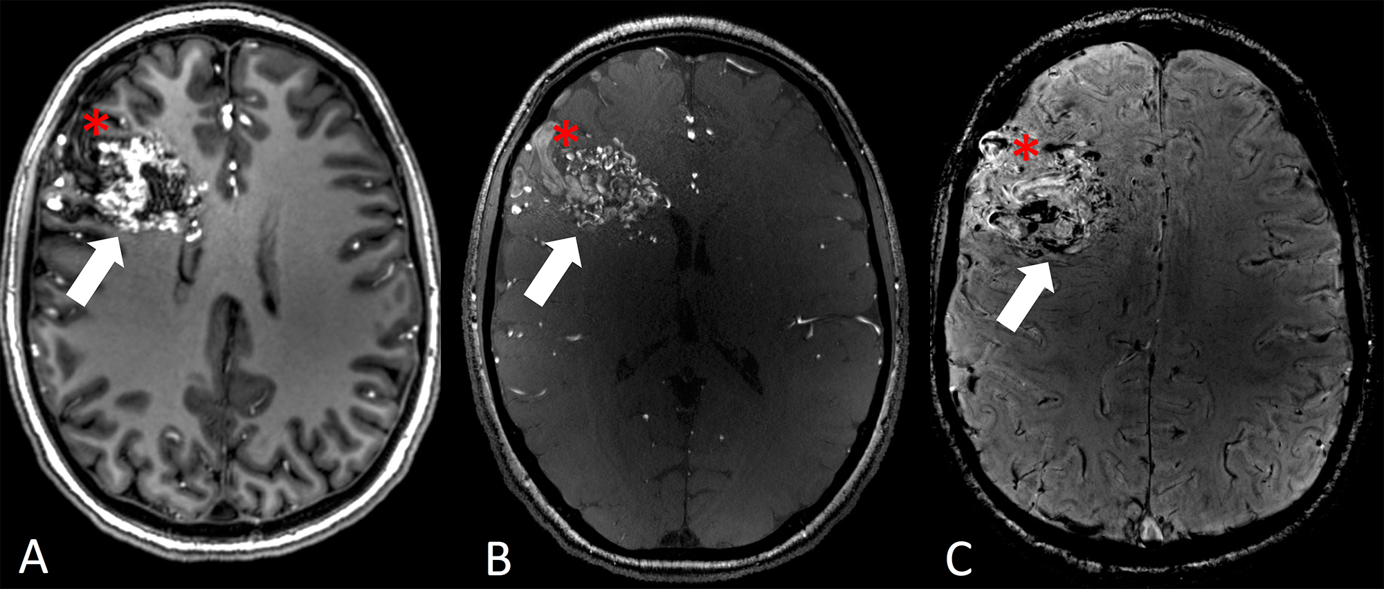
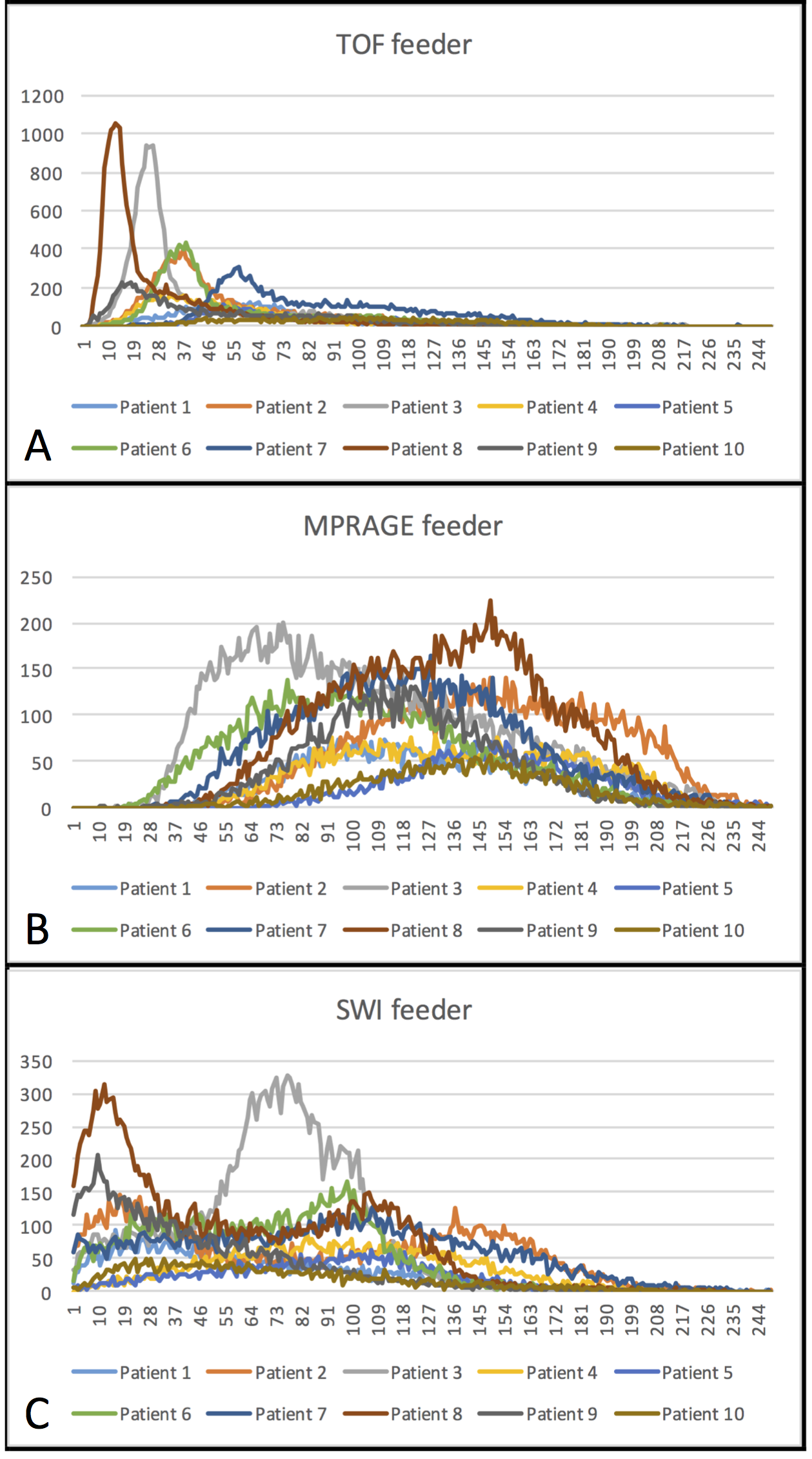
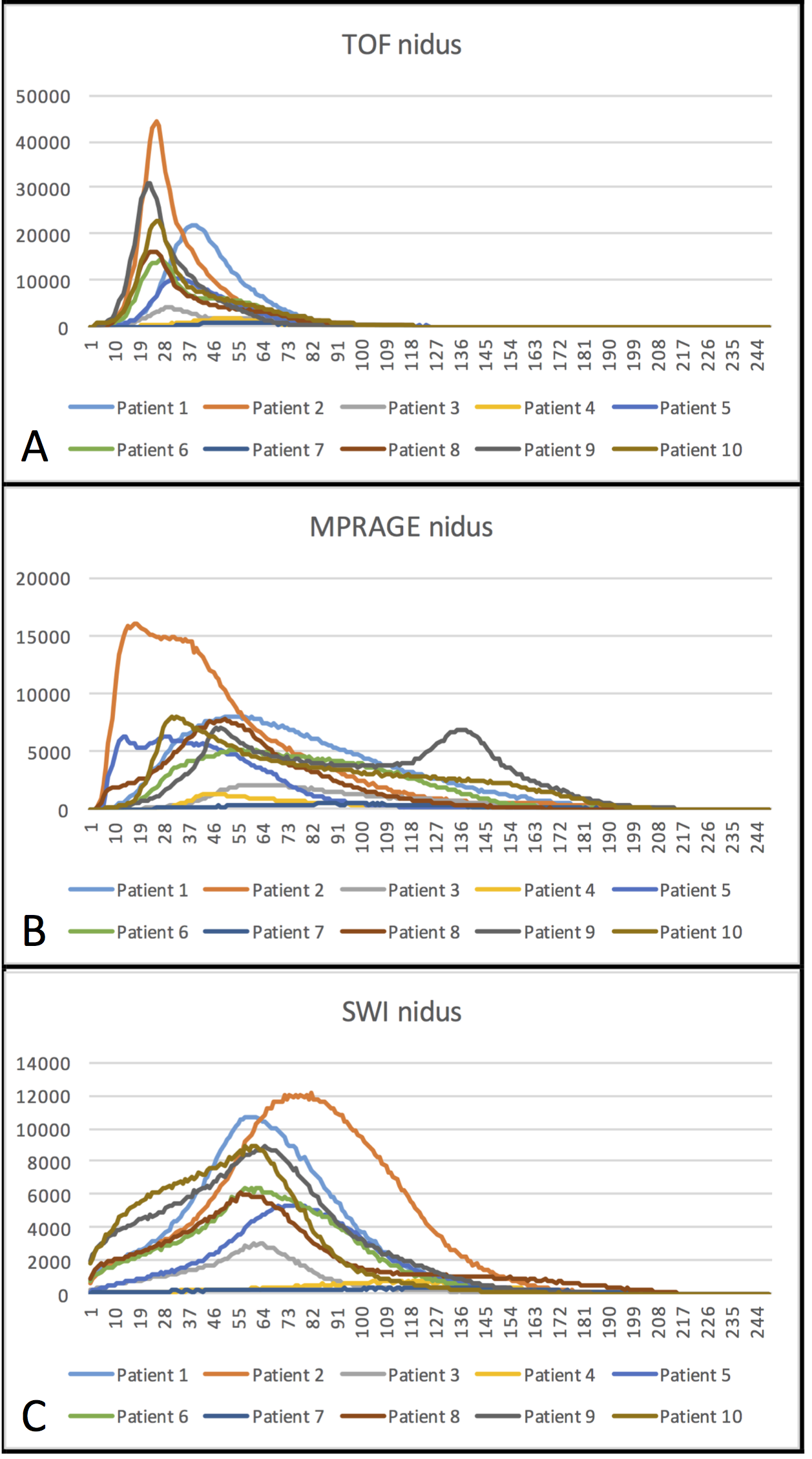
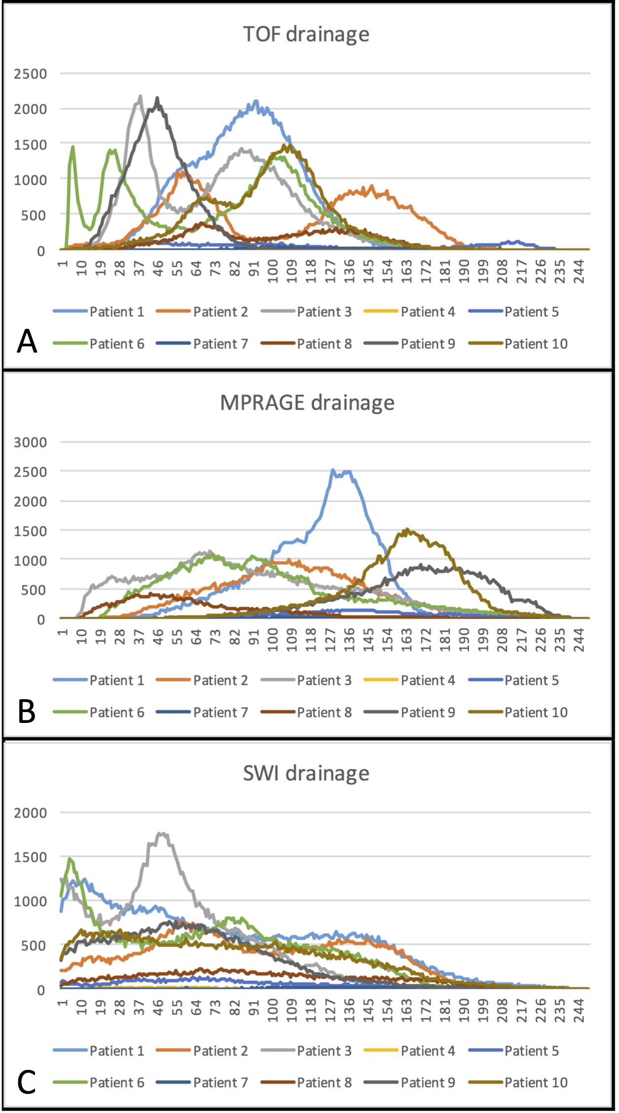
All examinations were successfully performed without occurrence of adverse events. Arterial AVM feeders could be identified excellently in TOF sequences. This correlates with narrow ranges of high signal intensities in the histograms (Figure 2 A). On the contrary, MPRAGE and SWI delineated AVM feeders heterogeneously. Correspondingly, histograms (Figure 2 B, 2 C) show a variable and wide signal intensity distribution.Delineation of the AVM nidus was homogeneous in TOF, MPRAGE and SWI for the majority of patients. The histograms show homogeneous signal intensity distributions, especially in the TOF sequences. (Figure 3 A-C). The venous drainage appeared inhomogeneous in all acquired sequences with hyperintense as well as hypointense findings. Intensity histograms reflect these results with variable peaks at all intensity levels as shown in Figure 4 A-C.
Discussion and Conclusion
As previously shown, 7 T UHF MRI has excellent imaging results regarding vessel delineation in AVMs. However, in contrast to conventional examinations (DSA and 3 T MRI) that depict arterial feeders, AVM nidus and venous drainage homogeneously, identification of the AVM architecture remains challenging in UHF 7 T MRI. Due to the signal heterogeneity especially in the feeders and drainage these structures might be misinterpreted in some cases. Furthermore, automated segmentation of AVMs depicted by 7 T UHF MRI does not seem feasible.Acknowledgements
The research leading to these results has received funding from the Interne Forschungsförderung Essen (IFORES), University Hospital Essen, University Duisburg-Essen.References
1. Mohr JP, Parides MK, Stapf C, et al. Medical management with or without interventional therapy for unruptured brain arteriovenous malformations (ARUBA): a multicentre, non-blinded, randomised trial. Lancet. 2014;383(9917):614-21.
2. Wrede KH, Dammann P, Johst S, et al. Non-Enhanced MR Imaging of Cerebral Arteriovenous Malformations at 7 Tesla. Eur Radiol. 2015 Jun 17.
3. Johst S, Wrede KH, Ladd ME, Maderwald S. Time-of-Flight Magnetic Resonance Angiography at 7 T Using Venous Saturation Pulses With Reduced Flip Angles. Invest Radiol. 2012 Aug;47(8):445-50.
4. Wrede KH, Johst S, Dammann P, et al. Caudal Image Contrast Inversion in MPRAGE at 7 Tesla Problem and Solution. Acad Radiol. 2012 Feb;19(2):172-8.
5. Dammann P, Kraff O, Wrede KH, et al. Evaluation of hardware-related geometrical distortion in structural MRI at 7 Tesla for image-guided applications in neurosurgery. Acad Radiol. 2011 Jul;18(7):910-6.
Figures