4409
Characteristics of fat in orbits of patients with early-stage hyperthyroidism1Ningxia people's hospital, yinchuan, People's Republic of China, 2Medical Imaging center, Ningxia people's hospital, People's Republic of China, 3Philips HealthCare, Beijing,china, People's Republic of China, 4People's Republic of China
Synopsis
Summary: Exophthalmos caused by muscle thickening and fat increasing in orbit is the typical symptom in hyperthyroidism, which impairs multiple systems as the auto-immune disease. From this study, we found the increase of percentage of fat and T2* value using the fat quantification analysis package, which indicate the accumulation and differentiation of fat-oriented cells, inflammation cells infiltration and collagen mucous change. Therefore, the characteristics of fat change in orbit can be evaluated by the transverse six-echo proton density fat fraction (PDFF) sequence (mDIXON-quant) technique in early-stage hyperthyroidism without significant manifestation of eyes.
Characteristics of fat in orbits of patients with early-stage hyperthyroidism
Introduction: Exophthalmos caused by muscle thickening and fat increasing in orbit is the typical symptom in hyperthyroidism, which impairs multiple systems as the auto-immune disease. Although there were lots of literature studied the morphology of orbit in hyperthyroidism by magnetic resonance imaging (MRI), research on quantification of fat in early-stage hyperthyroidism without significant exophthalmos is rare.
Purpose: To explore the characteristics of fat in orbits of early-phase hyperthyroidism patients without obvious abnormality in eyes.
Methods: 15 patients (30 eyes) diagnosed as early-phase hyperthyroidism without significant exophthalmos were recruited and 15 normal subjects (30 eyes) were involved as the control. All subjects were scanned in a whole body 3T (Ingenia, Philips Healthcare, Best, the Netherlands) MR with a 16ch ds head coil. A transverse six-echo proton density fat fraction (PDFF) sequence (mDIXON-quant) was employed with following parameters: TE/TR = shortest/9 ms, FA = 3.0°, matrix = 228 * 278, slice thickness = 3mm, slices = 20, NSA = 2. the degree of exophthalmos (Figure 1)1, fat thickness was measured by calculating number of slices with fat. Fat fraction and T2* values of fat were measured by drawing ROIs on the maximum slice with fat .The t test was used to compare the differences in contiguous data, and χ2 test for the categorical data.
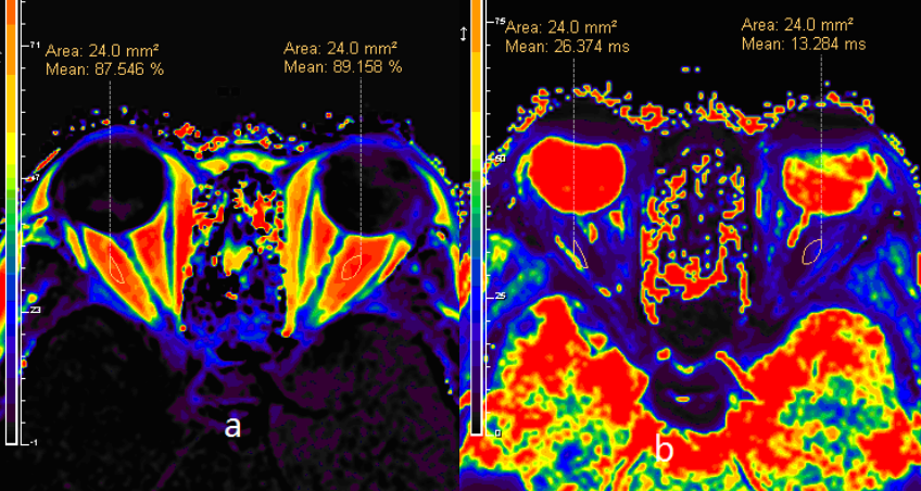
Results: 1.There was no significant differences in age between study and control groups (38.27 ± 9.96 vs. 37.80 ± 10.21 years, t = 0.127, P = 0.900 > 0.05), so was the sex (male 8 vs. 7 and female 7 vs. 8, χ2 = 0.133,P = 0.715 > 0.05). 2.The quantification of exophthalmos in study and control group were 16.10 ± 1.53 mm, 15.28 ± 1.99 mm, respectively. There was no significant difference (t = 1.746, p = 0.086﹥0.05).3. The thickness of fat in orbit in study and control group were 4.29 ± 0.54 mm, 4.06 ± 0.54 mm, respectively. There was no significant difference (t = 1.681, p = 0.099﹥0.05).4. The percentage of fat in orbit in study and control group were 87.96 ± 4.14 %, 83.75 ± 5.58 %, respectively. There was significant difference (t = 3.23, p = 0.002 < 0.05). The T2* value in study and control group were 28.31 ± 23.03 ms, 17.77 ± 10.11 ms, respectively. There was significant difference (t = 2.159, p = 0.035﹤0.05). (Table 1)(Figure 2. Figure 3)
Discussion: Fat in orbit increased in the early-stage of hyperthyroidism without remarkable manifestation, which signify the accumulation and differentiation of fat-oriented cells. Meanwhile, inflammation cells infiltration and collagen mucous change may cause the rise of T2* value.2
Conclusion: Orbits with early-stage hyperthyroidism have increased fat fraction and T2* value. The PDFF technique can be employed to diagnose the early-stage hyperthyroidism.
Acknowledgements
There is no acknowledgement.References
1.Comerci M, Elefante A, Strianese D, et al. Semiautomatic regional segmentation to measure orbital fat volumes in thyroid-associated ophthalmopathy. A validation study. Neuroradiol J. 2013; 26(4):373-379.
2.Garrity J, Bahn R. Pathogenesis of graves ophthalmopathy: implications for prediction, prevention, and treatment. Am J Ophthalmol. 2006; 142(1):147-153.
Figures
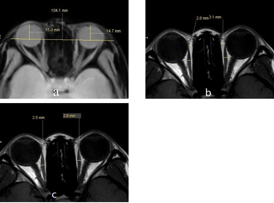
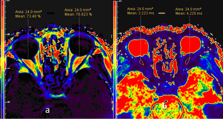
Figure 2: It shows the image of orbit in the normal patient. Both images have the same ROI area. a) The percentage of fat in right and left orbit is 73.48 % and 76.92%, respectively. b) The T2* value in right and left orbit is 3.22 and 4.22 ms, respectively.