4398
Optimization of b-values in iShim diffusion-weighted MR imaging of the thyroid gland: to improve the visualization of malignant thyroid nodules and the accuracy in differentiating malignant from benign thyroid nodulesQingjun Wang1, Qinglei Shi2, Tianyi Qian3, Alto Stemmer4, and Yong Guo1
1Department of Radiology, Chinese Navy General Hospital, Beijing, People's Republic of China, 2Scientific Marketing, Siemens Healthcare, People's Republic of China, 3MR Collaobration, HC NEA MR COL, Siemens Healthcare, People's Republic of China, 4MR Application Predevelopment, Siemens Healthcare GmbH
Synopsis
To investigate the
optimal b-values of diffusion-weighted imaging (DWI), a prototype sequence with
integrated slice-specific shimming (iShim) was applied for visualizing thyroid malignant
nodules and making accurate differentiation between benign and malignant
nodules. DWI images were acquired with five b-values including 0, 800, 1000,
2000 and 3000 sec/mm2. The results show that the iShim-DWI with b-value of 800
s/mm2 can present the clearest delineation of malignant thyroid
nodules and that the images with b-value of 3000 s/mm2 had the best
performances in differentiating malignant from benign thyroid nodules.
INTRODUCTION:
At the beginning the prototype sequence acquires a 2D-field map for each EPI imaging slice. The field-map is used to determine the optimal center frequency and gradient offsets for the imaging slice. Center frequency and gradient offsets are then updated before the acquisition of each imaging slice in real time. This sequence integrated shimming (iShim) technique is an effective method which could reduce the susceptibility artifacts at 3T for diffusion weighted imaging (DWI) and it also provide great potential for higher b-values imaging, especially in the neck and spine applications, as supported by the apparent improvement in signal integrity, spatial alignment.1 The purpose of this study was to determine the utility of iShim-DWI for visualizing thyroid malignant nodules and select the best b-values for differentiating malignant from benign thyroid nodules.MATERIALS AND METHODS:
Fifty-three benign thyroid nodules in 40 patients (7 men and 33 women, mean age 48.63±12.35 years) and 59 malignant thyroid nodules in 35 patients (8 men and 27 women, mean age 48.48±13.29 years), both of which were confirmed by pathology, underwent DWI exam using the prototype ishim sequence with 5 b-values (0, 800, 1000, 2000 and 3000 s/mm2) on a MAGNETOM Skyra 3T MR scanner (Siemens Healthcare, Erlangen, Germany). The images of b-values 800, 1000, 2000 and 3000 s/mm2 were retrospectively evaluated. Therefore the findings of thyroid malignant nodules were rated on a 3-point scale (type1 = clear hyperintensity relative to the surrounding normal thyroid gland, type2 = hyperintensity with an unclear distal border, and type3 = isointensity) and the subjective image quality of thyroid malignant nodules was rated on a 4-point scale{subjective scoring:4 = excellent (no problems were noticed in the image and the nodule was clearly shown), 3 = good (the image suffered from only minor degradation and was suitable for the evaluation of the nodule), 2 = moderate (image quality was not good but usable for the evaluation of the nodule), and 1 = poor (image quality precluded assessment of the nodule and the thyroid gland was barely shown)}. The iShim-DWI findings, subjective image quality score and the nodule-to-thyroid signal intensity (SI) ratios were measured and then compared among the 4 b-values using One-way ANOVA and Kruskal-Wallis One-way ANOVA with Bonferroni correction for post-hoc analysis. For the diagnostic ability of the nodule-to-thyroid SI ratios, receiver operating characteristic (ROC) analysis was conducted to assess the cutoff values and diagnostic performance.RESULTS:
There was a significantly higher incidence of malignant thyroid nodules showing clear hyperintensity on iShim-DWI with b = 800 s/mm2 than on the images with b-values of 1000, 2000 and 3000 s/mm2(P<0.01). The image quality was decreased when b-values increased from 800 to 3000 s/mm2 (P< 0.001). The lowest subjective scores of image quality were observed in b-value of 3000 s/mm2. However, there was highest nodule-to-thyroid SI ratio (2.25 ± 0.27) for malignant nodules and lowest nodule-to-thyroid SI ratio (0.64 ± 0.15) for benign nodules with b-value of 3000 s/mm2. In differentiating malignant from benign nodules, ROC analysis demonstrated a highest sensitivity, specificity and AUC on iShim-DWI with b-value of 3000 s/mm2 (90.62%, 93.16% and 0.96, respectively), followed by b = 2000 s/mm2 (85.71%, 87.50% and 0.90), 1000 s/mm2 (83.08%, 78.29% and 0.84) and 800 s/mm2 (81.38%, 75.00% and 0.82). The Fig. 1 and Fig. 2 demonstrated the difference in nodule-to-thyroid SI ratio on iShim-DWI images among different b-values between malignant and benign thyroid nodules.DISCUSSION & CONCLUSION:
Our results show that the images of b-value = 800 s/mm2 acquired with the iShim-DWI technique can improve the delineation of malignant thyroid nodules. Further they show that the images with b = 3000 s/mm2 can improve the diagnostic accuracy of differentiation between benign and malignant thyroid nodules better than other lower b-value images. So, we suggest that besides the b-value of 0 s/mm2, the other two b-values of 800 and 3000 s/mm2 should be the optimal b-values for DWI in thyroid gland applications.Acknowledgements
No acknowledgement found.References
1. Zhang H, Xue H, Alto S, et al. Integrated shimming improves lesion detection in whole-body diffusion-weighted examinations of patients with plasma disorder at 3T. Invest Radiol. 2016; 51(5): 297-305.Figures
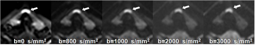
Fig. 1 A papillary
carcinoma in the isthmus of thyroid gland in a 36-year-old woman. The iShim-DWI
with b-value of 800 s/mm2 showed most clear margin of the nodule and the image with
b-value of 3000 s/mm2 showed the highest nodule-to-thyroid SI ratio of the
nodule (white arrows).
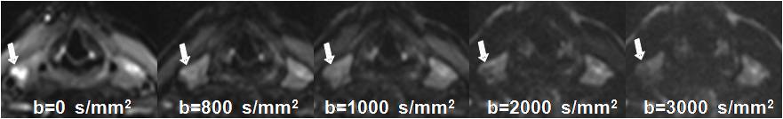
Fig. 2 A goiter in the
right lobe in a 45-year-old man. The signal intensity of the nodule decreased
with increasing b-values and the image with b-value of 3000 s/mm2 showed the
lowest nodule-to-thyroid SI ratio of the nodule (white arrows).