4381
Arterial Input Function Selection in DSC-MRI of Brain Tumors Using Differential Evaluation Clustering MethodHossein Rahim Zadeh1, Anahita Fathi Kazerooni1,2, Mohammad Reza Deevband3, and Hamidreza Saligheh Rad1
1Quantitative MR Imaging and Spectroscopy Group, Research Center for Molecular and Cellular Imaging, Tehran University of Medical Sciences, Tehran, Iran, 2Department of Medical Physics and Biomedical Engineering, Tehran University of Medical Sciences, Tehran, Iran, 3Shahid Beheshti University of Medical Sciences, Tehran, Iran
Synopsis
Proper arterial input function (AIF) selection is a critical step for accurate quantification of dynamic susceptibility contrast enhanced (DSC) MRI in brain tumor patients. In this study, we have employed differential evaluation (DE) clustering method on processed perfusion images for accurate AIF selection. The procedure consists of two main steps: preprocessing for eliminating non-arterial curves including tissue, noisy and those contaminated with partial volume effects; and AIF selection using DE clustering method. The performance of this clustering method was compared to K-means and Hierarchical Clustering techniques and the results show the superiority of the proposed approach for accurate AIF selection.
Introduction
Arterial Input Function (AIF) selection plays an important role in absolute quantification of perfusion parameters using dynamic susceptibility contrast enhanced (DSC-) MRI 1. Manual AIF selection is time-consuming and depends on the expertise of the radiologist. So, employing automatic methods is desirable. The main disadvantage of previously-proposed automatic AIF selection methods is their lack of information about the global shape of the clusters which complicates separation of overlapping clusters, and their implementation on normal subjects; therefore, their performance can be compromised in cases of analysis for brain tumor patients 2. Hence, in this study, we evaluated metaheuristic differential evolution (DE) algorithm to find a global optimal solution for optimization of clusters during the search for the proper AIF.Methods
In this study, DSC-MR images of 43 patients (20 low-grade glioma, and 23 glioblastoma multiforme (GBM)) with brain tumors were downloaded from the Cancer Imaging Archive Database 3. Automatic AIF selection depends on the characteristics of concentration-time curve, such as the maximum peak (MP), full width at half maximum (FWHM), area under curve (AUC), time to peak (TTP). Arterial curve has higher maximum peak, area under curve and lower time to peak, FWHM in comparison others. The steps for AIF selection are as follow: (1) consecutive perfusion images in a series were aligned to the first pre-contrast image; (2) to eliminate the curves of tumorous region that mimic arterial curves, k-means clustering was used to classify the signal intensity curves into five clusters. As the mean signal intensity of the baseline for the tumorous region is higher than other brain regions, the cluster with the highest mean baseline value was removed as a tumorous cluster; (3) area under the concentration curves was calculated and 90% of the curves with lowest values were discarded; (4) Differential evolution algorithm with 5 clusters was applied to the remaining curves AIF cluster was selected by calculating the measure M=MP/ (TTP × FWHM). The performance of this method in AIF selection according to the expert’s selection was compared against K-means and Agglomerative Hierarchical (AH) clustering methods.Results
Average value of automatic and manual AIF shape parameters i.e. FWHM, time to peak, maximum peak, area under curve and M -value (M=MP/ (TTP × FWHM)) were calculated for 43 data and used for statistical analysis. Then, the differences between automatic and manual AIF were computed by root-mean-square (RMS). Statistical analysis using paired t-test was used (with significance level of P-value<0.05). The mean values of AIF shape parameters obtained by AH and DE clustering were closer to manual selection and RMSE decreased remarkably in comparison with K-means clustering. Among AH and DE clustering methods, DE showed lower RMSE error and mean values of AIF shape parameters (Table1) in DE clustering showed better results.Discussion
Finding proper clustering method for AIF selection has been challenging. So, in this study, we evaluated the capability of differential evaluation clustering method for AIF selection. According to Table 1, the mean values and paired t-test analysis results, both AH and DE clustering methods had better result in comparison K-means clustering. In RMSE measurement, DE outperformed AH, meaning that the chosen AIF by DE clustering method was closer to manual selection in comparison with AH method. In some data, all three clustering methods, wrongly selected noisy and truncated curves as AIF (Figs.1, 2).Conclusion
The proposed pre-processing and clustering approach for AIF selection, showed good agreement with the AIFs selected by an expert, implying that it is necessary to carefully undertake preprocessing steps besides employing an accurate clustering method, to increase the accuracy and precision of automatic AIF selection.Acknowledgements
No acknowledgement found.References
1. Shiroishi MS, Castellazzi G, Boxerman JL, D'Amore F, Essig M, Nguyen TB, et al. Principles of T2*-weighted dynamic susceptibility contrast MRI technique in brain tumor imaging. Journal of Magnetic Resonance Imaging. 2015;41(2):296-313. 2. Das S, Abraham A, Konar A. Automatic clustering using an improved differential evolution algorithm. IEEE Transactions on systems, man, and cybernetics-Part A: Systems and Humans. 2008;38(1):218-37. 3. Clark K, Vendt B, Smith K, Freymann J, Kirby J, Koppel P, et al. The Cancer Imaging Archive (TCIA): maintaining and operating a public information repository. Journal of digital imaging. 2013;26(6):1045-57.Figures
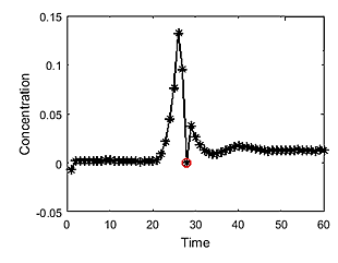
FIG. 1. An example of a truncated concentration curve wrongly chosen as
AIF
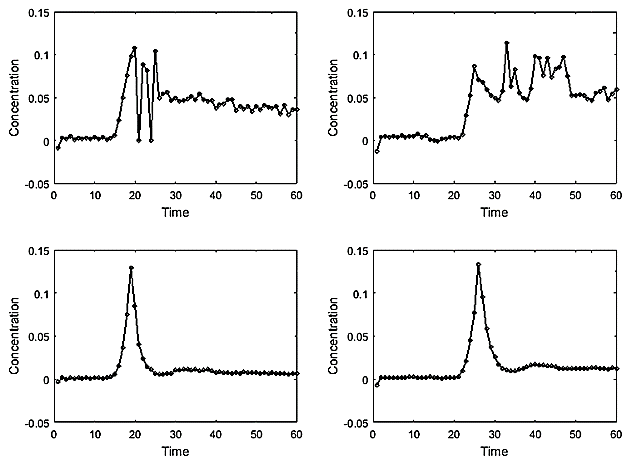
FIG. 2. An example of noisy concentration curves wrongly chosen as AIF
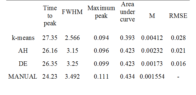
Table 1. Comparison of the mean values of AIF shape parameters obtained
using different clustering approaches (DE, AH, and K-means)