4371
Near infrared photoimmunotherapy for lung cancer in a transgenic mouse model evaluated by MRI1National Cancer Institute, Bethesda, MD, United States, 2Diagnostic Radiology, Hiroshima University, Hiroshima, Japan, 3Frederick National Laboratory for Cancer Research
Synopsis
Near infrared photoimmunotherapy (NIR-PIT) is a new cancer treatment that combines the specificity of antibodies for targeting tumors with the toxicity induced by photoabsorbers after irradiation with NIR light. The purpose of this study was to determine whether MRI can monitor the therapeutic effect of NIR-PIT in spontaneously occurring lung cancers that express epidermal growth factor receptor. Tumor volume ratio was inhibited significantly in the NIR-PIT group compared with control group. Thus, MRI can be a useful imaging modality for monitoring the therapeutic effects of NIR-PIT for cancer.
Introduction
Near infrared photoimmunotherapy (NIR-PIT) is a new cancer treatment that combines the specificity of antibodies for targeting tumors with the toxicity induced by photoabsorbers after irradiation with NIR light1. In xenografts the therapeutic effect of NIR-PIT can be measured with a caliper or luciferase activity 1-4. However, measurement of tumor size with a caliper is available only for subcutaneous tumors, and the tumor cells must be previously transfected with the luciferase gene for bioluminescence. On the other hand, magnetic resonance imaging (MRI) is a widely used imaging modality that depicts both anatomic and functional information in a short time with high tissue contrast. The advantages of MRI include whole body scanning regardless of the depth and that it does not require transfection of luciferase or contrast agents. NIR-PIT has been demonstrated to be effective using subcutaneously xenografted human tumors in athymic mice. However, cancers growing in transgenic mouse models are more similar to cancers in patients5. The purpose of this study was to monitor with MRI the therapeutic effect of NIR-PIT in a transgenic model with spontaneously occurring lung cancer which expresses human epidermal growth factor receptor (hEGFR).Methods
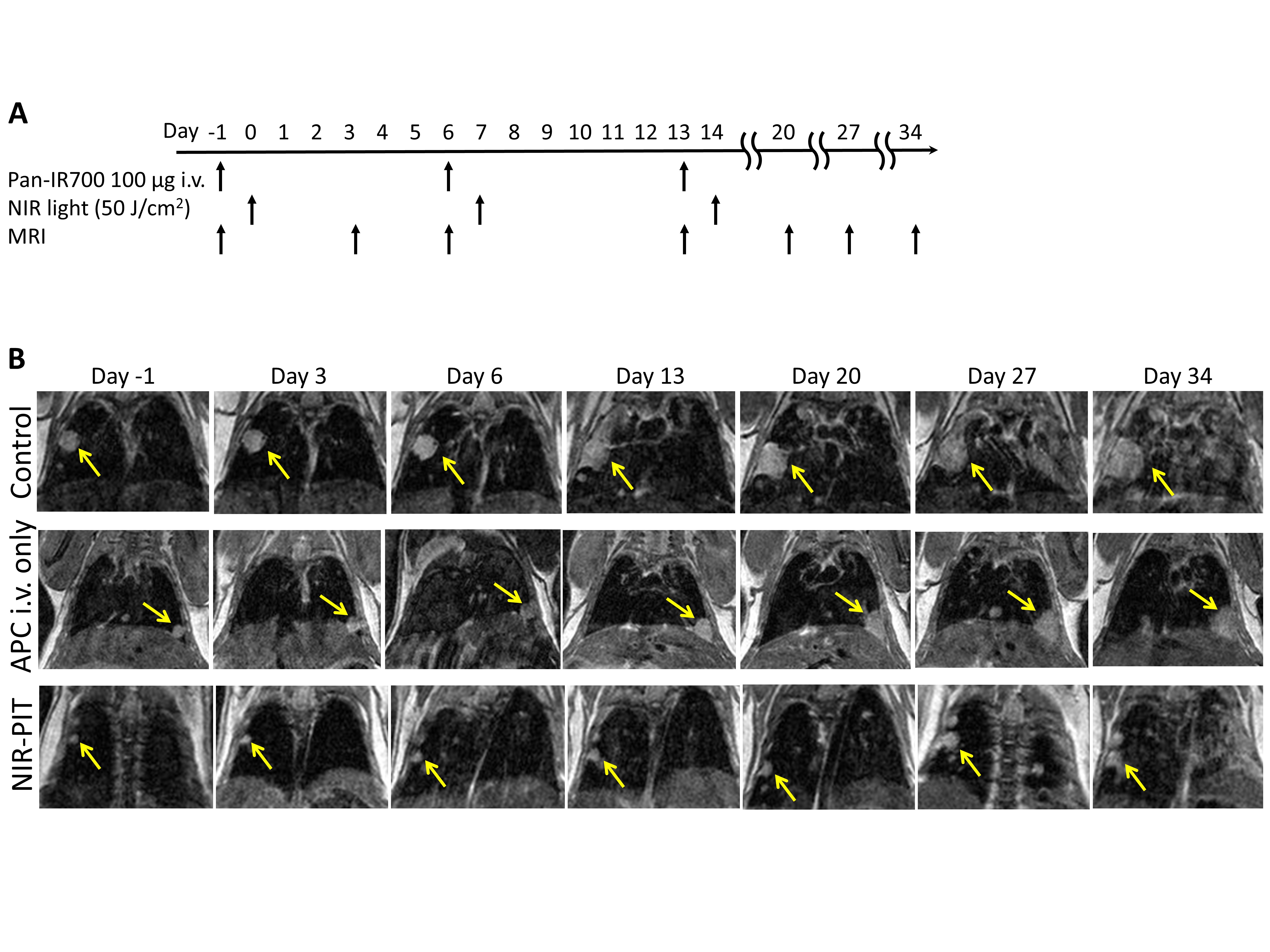
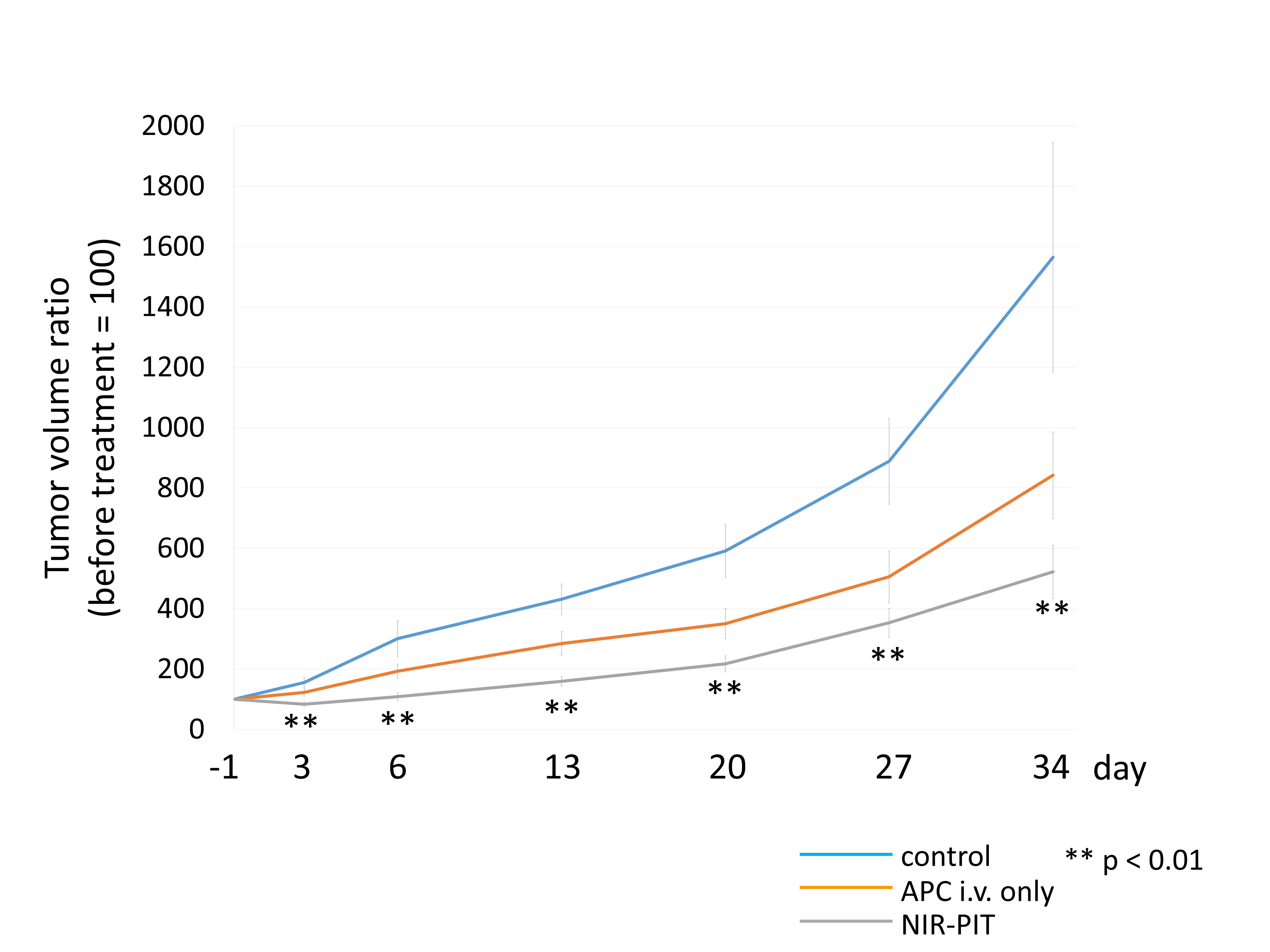
We used the hEGFR TL transgenic mice which produce hEGFR expressing lung cancers spontaneously. Mice were separated into 3 groups for the following treatments: (1) no treatment (control); (2) 150 μg of photoabsorber, IR700, conjugated to panitumumab, an antibody targeting EGFR (antibody-photoabsorber conjugate (APC)) i.v. only (no NIR light) ; (3) 150 μg of APC i.v. and NIR light administered from 2 directions (each 25 J/cm2) via the back and front on day 1. Each treatment was performed every week up to three weeks. All mice underwent T2-weighted imaging (T2-WI), T1-weighted spoiled gradient-echo sequence, and balanced-steady-state free precession (b-FFE). The volume of the lung tumors was monitored by MRI and a tumor volume ratio was determined at each time point relative to the initial tumor volume. Up to 5 of the largest lesions were evaluated when multiple lesions were present. Steel’s test for multiple comparison was used to compare the tumor volume ratio with that of control. Differences of p < 0.05 were considered statistically significant.Results
The treatment regimen including MR imaging is shown in Figure 1A. In the control and APC i.v. only groups the size of lung tumor increased rapidly consistent with the known growth rates of these tumors. In the NIR-PIT treatment group the size of lung tumors also continued to increase but at a much slower rate (Figure 1B). Tumor volume ratios were significantly lower in the NIR-PIT group compared with control group (p < 0.01 at all time points). No significant difference in the tumor volume ratio was observed between the APC i.v. only group and the control group (p = 0.43, 0.34, 0.09, 0.12, 0.19, and 0.45 at day 3, 6, 13, 20, 27, and 34, respectively) (Figure 2).Discussion
The tumor volume ratio measured by MRI was reduced significantly by treatment of these spontaneously occurring lung tumors only in the NIR-PIT treatment group. The therapeutic effect was clearly visible on serial MRIs. Previous experiments with NIR-PIT using HER2-expressing xenografted lung metastases in athymic nude mice evaluated by luciferase activity showed a greater effect than NIR-PIT for lung cancer in transgenic mice model evaluated by MRI 4. This may be because MRI can evaluate only morphological change, while luciferase activity can detect early necrotic cell death preceding the morphological change. However, MRI is much more widely available, requires no transfection, is useful in all parts of the body and does not expose the subject to ionizing radiation. Therefore, this result suggests that MRI is useful, if not as sensitive as bioluminescence in detecting response to NIR-PIT in the lungs.Conclusion
Tumor volume ratios measured by MRI were inhibited significantly by NIR-PIT treatment compared with a control group in a transgenic model of lung cancer. NIR-PIT was effective in this transgenic mouse model and MRI was useful as an imaging modality for monitoring the therapeutic effect of NIR-PIT for cancers.Acknowledgements
No acknowledgement found.References
1. Mitsunaga M, Ogawa M, Kosaka N, et al. Cancer cell-selective in vivo near infrared photoimmunotherapy targeting specific membrane molecules. Nature medicine. 2011;17(12):1685-91.
2. Rehemtulla A, Stegman LD, Cardozo SJ, et al. Rapid and quantitative assessment of cancer treatment response using in vivo bioluminescence imaging. Neoplasia. 2000;2(6):491-5.
3. Sato K, Nakajima T, Choyke PL, et al. Selective cell elimination in vitro and in vivo from tissues and tumors using antibodies conjugated with a near infrared phthalocyanine. RSC advances. 2015;5(32):25105-14.
4. Sato K, Nagaya T, Mitsunaga M, et al. Near infrared photoimmunotherapy for lung metastases. Cancer Lett. 2015;365(1):112-21.
5. Hoffman RM. Patient-derived orthotopic xenografts: better mimic of metastasis than subcutaneous xenografts. Nature reviews Cancer. 2015;15(8):451-2.
Figures