4349
Unified Coils (UNIC) for Simultaneous RF Reception and Targeted Local B0 Shimming1Biomedical Imaging Research Institute, Cedars-Sinai Medical Center, LOS ANGELES, CA, United States
Synopsis
We propose a new general MR coil concept with integrated RF and Bo shimming applicable to almost all MRI coils and systems. Innovative geometrical decoupling methods are proposed to bring the distance between separate shim and RF loops to zero millimeters and to make shim loops physically free from the RF loops. Therefore, both RF and shim coils can be in close proximity to the subject and be designed independently to maximize the performance of each function. It also opens a new window to integrate shim and RF coils or other coils in the constricted scanner bore space.
INTRODUCTION
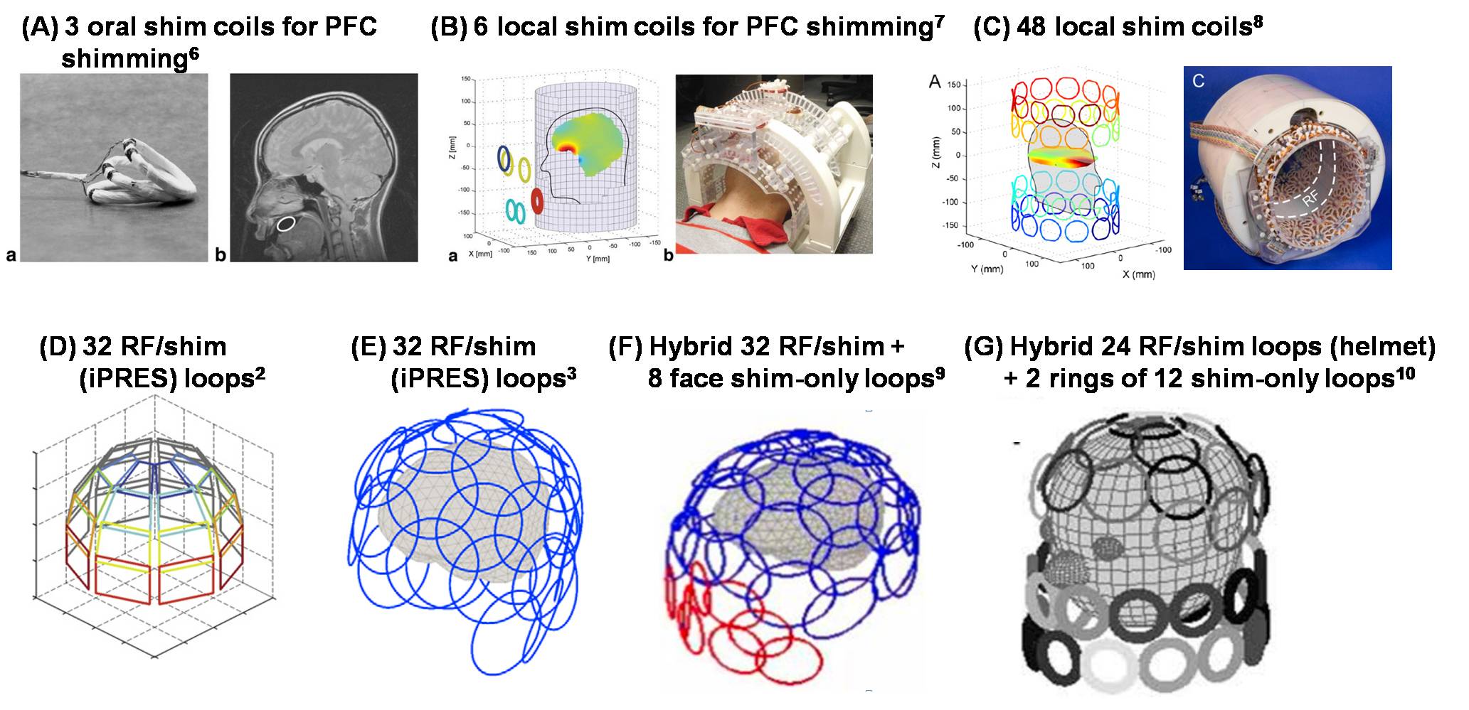
To propose a new general MR coil concept with integrated B0 shimming applicable to almost all MR coils and imaging systems. As one example of many off-resonance imaging problems, image artifacts and signal voids compromise whole brain fMRI particularly in prefrontal cortex (PFC) and temporal lobes (TLs). Previously, we proposed a so-called iPRES1 concept using the same single coil array for simultaneous B0 shimming and RF reception by adding DC currents to RF reception coil loops.1-4 It has drawn great attention. The dual shim-RF functions in one loop, however, also poses inherent limitations: i) The design of shim loops (size, shape, and position) restricted to that of the corresponding RF loops. ii) The total number of shim loops (typically 32 or less) and loop turns (only 1-turn) also were constrained. iii) Many large-size RF chokes required to reduce RF SNR loss, complicating coil construction and problematic at 7T.5 Due to these limitations its capability and flexibility in local shimming are largely undermined. Fig.1 overviews state-of-the-art designs.2-3,6-10 Here we propose a new concept 'UNIC' for unified coils to well solve these limits and largely improve field homogeneity. Moreover, unified coil assembly can remain the same dimensions compared to clinical RF coils.11METHODS
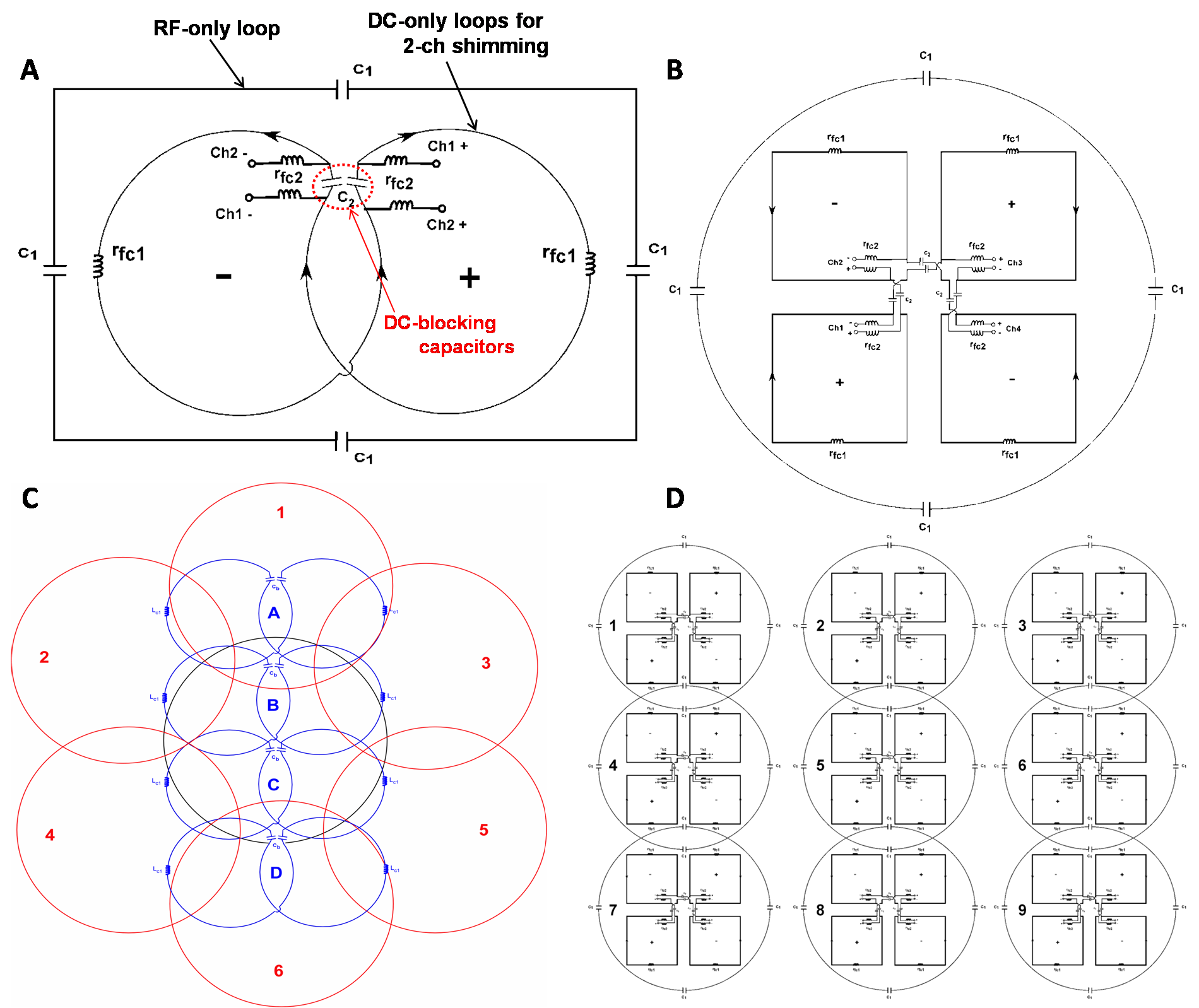
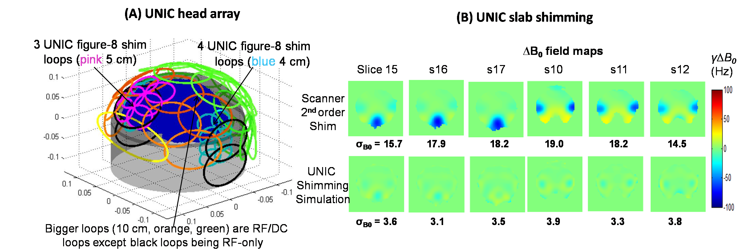
Fig.2A explains the idea and depicts the typical element design. The outer rectangular loop is a traditional RF-only loop. Inner two circular loops are 2-channel shim-only DC loops, forming a figure-8 shape by connecting two DC-blocking capacitors C2. While RF currents would pass C2 and follow figure-8 pathway (arrows), DC currents are restricted (by C2) to two separate circular shim loops, each independently driven by one current source (ch1+/-, ch2+/-). Geometrical symmetry ensures that undesired RF currents induced by the RF loop would create magnetic fluxes in two shim loops having equal magnitude but opposite polarities (+ -), thus cancelling out. Shim and RF loops have zero mutual inductance (RF-decoupled). The idea can be extended to 2N shim loops (Fig.1B). Critical overlap geometrical decoupling can also null mutual inductance as proven in Fig.3D. Roemer's pioneering work11 suggests a wide range of overlapping ratios can greatly reduce coupling. Fig.2C shows an array design for brain imaging. Red loops are RF/DC shared-conductor loops (iPRES) while blue loops are four separate figure-8 shim loops targeting PFC or TLs shimming. Figure-8 shim loops (ABCD) are inherently decoupled from both the black loop and red loops #1#6 as explained in Fig.2A and largely decoupled from red loops #2-5 by partial-overlap decoupling. Residual coupling can be eliminated by few RF chokes. The entire coil design is shown in Fig.4A, where sizes of figure-8 loops match those of targeted anatomical structures. In addition, separate shim loops enable multiple-turn, overcoming another major limit of iPRES loops in shim field strength.RESULTS
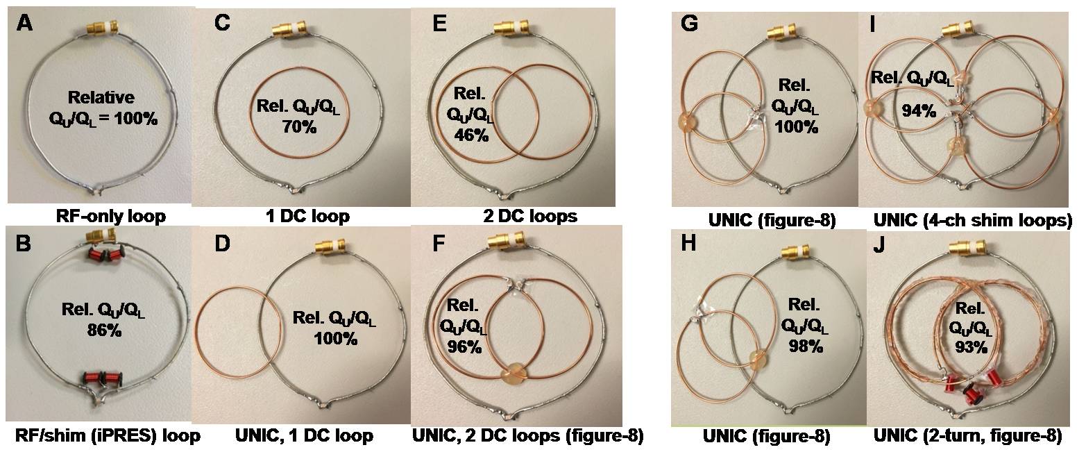
For bench measurement in Fig.3, without proposed geometrical decoupling, one and two separate shim loops as used in,5 experienced dramatic drops in QUnloaded/QLoaded, to 70% (C) and 46% (E) compared to RF-only loop (A). With UNIC, 1-turn figure-8 loop (for 2-ch shimming) only dropped to 96% (F) and had no drop in (D) using critical overlapping decoupling. Neighboring coupling were also negligible (G,H). Based on Fig.2B, the butterfly shape design (I) with 4-ch shim loops only had a Q-ratio drop 94%. Note all (Figs.3F-I) have no RF chokes but better Q-ratios than iPRES loop (B)! 2-turn (J) and 3-turn (not shown) figure-8 loops with 1-2 chokes per channel had slight drops 92-93%. Fig.4A shows UNIC head coil design (under construction). Simulation shows about 80% reduction in standard deviation of field offsets for all slabs in PFC and TLs, achieving unprecedented performance particularly for shimming TLs.DISCUSSION /CONCLUSION
The common major difficulty encountered when integrating shim with RF is the strong RF interaction between DC and RF pathways (Fig.1). We propose simple geometrical decoupling methods to bring the distance between separate shim and RF loops to zero millimeters and to make shim loops physically free from the RF loops. Therefore, both RF and shim coils can be in close proximity to the subject and be designed independently to maximize the performance of each function. Moreover, new coils can be potentially used in clinics without increasing patient discomfort. Simulation demonstrates new coil can reduce brain field inhomogeneity dramatically to meet the unmet challenge in fMRI of a real whole brain. New designs are applicable to almost all MR coils and systems including coils with less receivers (1-8), birdcage coils, and even animal scanners to meet various imaging challenges from head to toe with fMRI, DTI, MRSI, and etc.Acknowledgements
Thanks to Fraser Robb, Miguel Navarro, Bernd Stoeckel, Xiaoming Bi for their support.References
1. Han H, Song AW, Truong TK. Integrated parallel reception, excitation, and shimming (iPRES). Magn Reson Med 2013;70:241–247.
2.Truong TK, Darnell D, Song AW. Integrated RF/shim coil array for parallel reception and localized B0 shimming in the human brain. NeuroImage 2014;103:235–240.
3. Stockmann J, Witzel TP, Keil B, et al. A 32-channel combined RF and B0 shim array for 3T brain imaging. Magn Reson Med 2016;75:441–451.
4. Darnell D, Truong TK, Song AW. Integrated parallel reception, excitation, and shimming (iPRES) with multiple shim loops per radio-frequency coil element for improved B0 shimming Magn Reson Med 2016 DOI: 10.1002/mrm.26267.
5. Winkler SA, Stockmann JP, Warr PA, et al. Comparison of new element designs for combined RF-shim arrays at 7T. Proc. Intl. Soc. Mag. Reson. Med. 23 (2015) p0860.
6. Hsu JJ, Glover GH. Mitigation of susceptibility-induced signal loss in neuroimaging using localized shim coils. Magn Reson Med 2005;53:243–248.
7. Juchem C, Nixon TW, McIntyre S, et al. Magnetic field homogenization of the human prefrontal cortex with a set of localized electrical coils, Magn. Reson. Med. 2010;63:171–180.
8. Juchem C, Nixon TW, McIntyre S, et al. Dynamic multi-coil shimming of the human brain at 7T. J Magn Reson 2011;212:280–8.
9. Stockmann JP, Guerin B, Wald LL. Improving the efficiency of integrated RF-shim arrays using hybrid coil designs and channel placement and compression via a genetic algorithm Proc. Intl. Soc. Mag. Reson. Med. 23 (2016) p1153.
10. Juchem C, De Graaf RA. B0 Shimming Technology. Chapter 4 in 'Magnetic Resonance Technology: Hardware and System Component Design'. 'New Developments in NMR' No. 7. Published by the Royal Society of Chemistry, 2016.
11. Roemer PB, Edelstein WA, Hayes CE, et al. The NMR phased array. Magn Reson Med 1990;16:192-225.
Figures