4335
Very low field MRI setup for brain imaging1Commissariat à l'enegie Atomique, Saclay, France, Metropolitan
Synopsis
Mixed sensors coupled to tuned flux transformer could more effective than classical tuned coils in detecting MRI signals on a very low field range. In order to compare later on their effectiveness, first, a very low field head MRI system (8.4 mT) is developed. Homogeneity, gradients strength, excitation and reception coils were adjusted. A homogeneity of 84 ppm was achieved in a quasi-open configuration. The amplitude of each gradient was 100 times lower than at high field but sufficiently high to achieve a resolution of less 2 mm x 2 mm x 2 mm. Images with a 3D imaging acquisition without pre-polarization technique nor magnetic shielding room were achieved.
Introduction
Nowadays,
Ultra Low field (ULF) Magnetic Resonance Imaging (MRI), is getting more
attention for many reasons like the increased T1 contrast of tissues [1],
compatibility with patients having metallic implants and the possibility of
integrating magnetoencephalography studies. Several groups employed low Tc-SQUIDs
as a signal detector [2,3]. However, SQUIDs cannot handle strong magnetic field.
This limits the measurement field to few hundreds of µT. Thus, prepolariszation,
which is a time consuming method, is inevitable to increase polarization at
these range of field. Our laboratory has developed Nitrogen cooled
superconducting-magnetoresistive hybrid sensors [4] competitive to low Tc
SQUIDs above 100 kHz. These sensors are untuned and can be coupled through low
resistive room temperature flux transformers to a room temperature antenna
presenting a high filling factor. They are very robust and
accept strong RF pulses with a very short recovery time compared to tuned RF
coils, which allow measurements of broad signals. The efficiency of a mixed
sensor was proved on Very Low field (VLF) MRI setup with a FOV of 5x5x5cm3 [5].
The detectivity of the mixed sensor associated to a tuned flux transformer having
a pick up coil of 6 cm diameter is about 1.7 ft/Hz1/2
. In
order to carry on this technology into clinical, a whole
head VLF MRI system (FOV= 20x20x20
cm3) without pre-polarization technique nor magnetic
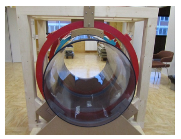
shielding room, is constructed (figure 1). Before setting mixed sensor within
the system, homogeneity, coils and gradients are developed and adjusted and 3D images
are performed in this workMethod
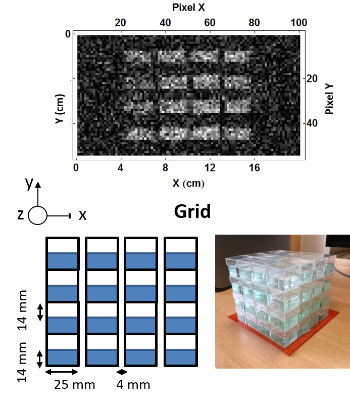
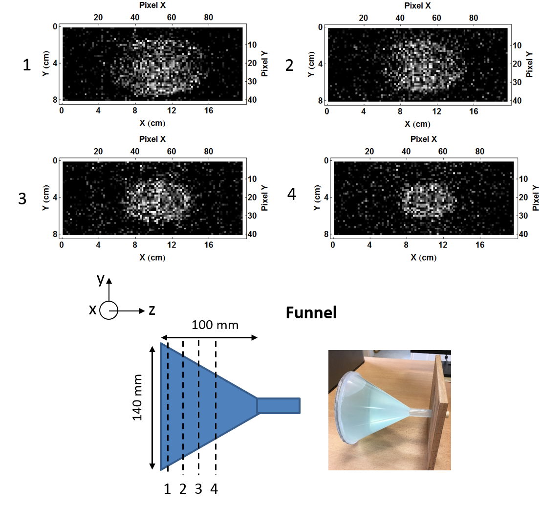
The main field, was set to 8.4 mT (359 kHz). The detector is a Helmholtz coil with dimensions corresponding to those head (10 cm radius).It is tuned to Larmor frequency with a quality factor of 60. The measured detectivity of this coil is about 10 1.7 ft/Hz1/2 at 359 kHz. Excitation coil is a saddle coil tuned at Larmor frequency and orientated perpendicular to the receiver coil in order to minimize coupling. Two different shapes of samples were imaged: A 3D grid of cubes (4 x 4 x 4 cubes) and a funnel. Samples are filled with doped water (T1 ≈ 130 ms; T2 ≈ 80 ms) simulating relaxation time of a brain. Gradient coils are used not only for 3D imaging but also for correcting external inhomogeneity (shimming). Achieved homogeneity is of 83 ppm corresponding to a resolution bandwidth of 30 Hz (1 point = 30 Hz). Thus, to achieve, for example, a resolution of 2 mm on a 20 cm length, sampling with at least 100 of points, in a given direction, is required. Consequently, gradient in that direction has to be set to 350 µT/m/A. A spin echo sequence was applied (TE=33 ms, TR=180 ms) with an acquisition bandwidth of 3 kHz.Results
First, one slice MRI image of the grid sample is performed (figure 2). A resolution of 2 x 2 mm2 on a FOV of 200 x 11 mm2 was achievable in a 5.8 minutes. Each point was averaged 35 times. SNR is about 5.
Then a 3D image acquisition is applied to get an image of the funnel sample (figure 3). A 2 x 4 x 12 mm3 resolution on a 200 x 160 x 50 mm3 FOV was achievable in a 14 minutes. Each point was also averaged 35 times and. SNR is about 3.
Discussion
We should note that those images, obtained with inductive Helmholtz coil, are raw images without any signal processing.
Knowing that the detectivity of a mixed sensor can reach less than 0.1 ft/Hz1/2 by enhancing flux transformer and enlarging the pick up coil diameter (10 cm diameter),100-fold gain in SNR is expected relatively to the currently used inductive coil, leading to images with a better SNR in a shorter time.
Conclusion
Developing and adjusting a brain VLF MRI system was done. Images of 200 x 160 x 50 mm3 dimensions and 2 x 4 x 12 mm3 resolution was achievable in 14 minutes using a full Cartesian 3D image acquisition. Future work will focus on reducing 3D imaging acquisition time by adopting gradient echo or Multi spin echo sequences and/or optimizing k space filling. Simultaneously, in-vivo experiments should be done. Then mixed sensors will be combined to the VLF MRI setup.Acknowledgements
We acknowledge the Technosanté program support.References
[1] Lee et al. Magn Reson Med. 53(1):9-14 (2005)
[2] Seton et al. Magn.Reson. Mat. In Physics, Biologie and Medecine 8 :116-120 (1999)
[3] Espy et al. J. Magn. Reson. 229:127-141 (2013)
[4] Pannetier et al. Science 304(5677):1648–1650 (2004)
[5] Herreros et al. Rev. Sci Instrum 84: 095116 (2013)
Figures