4331
A Compact Affordable Three-Bore Cryogen-Free Superconducting Magnet for Extremity ImagingShahin Pourrahimi1, Jerome L. Ackerman2,3, John E. C. William1, Nadder Pourrahimi1, and Alexey Kaplan1
1Superconducting Systems, Inc., Billerica, MA, United States, 2Martinos Center, Dept of Radiology, Massachusetts General Hospital, Charlestown, MA, United States, 3Department of Radiology, Harvard Medical School, Boston, MA, United States
Synopsis
In a project to develop a compact MRI limb scanner for orthopedic and metabolic bone disease applications we developed a three-bore 1.5T magnet with the following design goals: ability to operate the magnet in a small point-of-care space, elimination of liquid cryogens for installation or operation, a comfortable patient experience while scanning knees, ability to tilt the magnet to accommodate patients rather than requiring patients to accommodate to the magnet, and capability for conventional and solid state proton and phosphorus MRI for metabolic bone disease assessment.
Introduction
The overall goal of this project is to develop a compact MRI limb scanner for orthopedic and metabolic bone disease applications. The design goals included the ability to operate the magnet in a small point-of-care space, complete elimination of liquid cryogens for installation or operation, a comfortable patient experience while scanning knees, the ability to tilt the magnet to accommodate patients rather than requiring patients to accommodate to the magnet, and both conventional and solid state proton and phosphorus MRI for metabolic bone disease assessment. The following reports the results of the design and construction of a prototype cryogen-free three-bore superconducting 1.5T magnet as the first milestone in the project.Methods
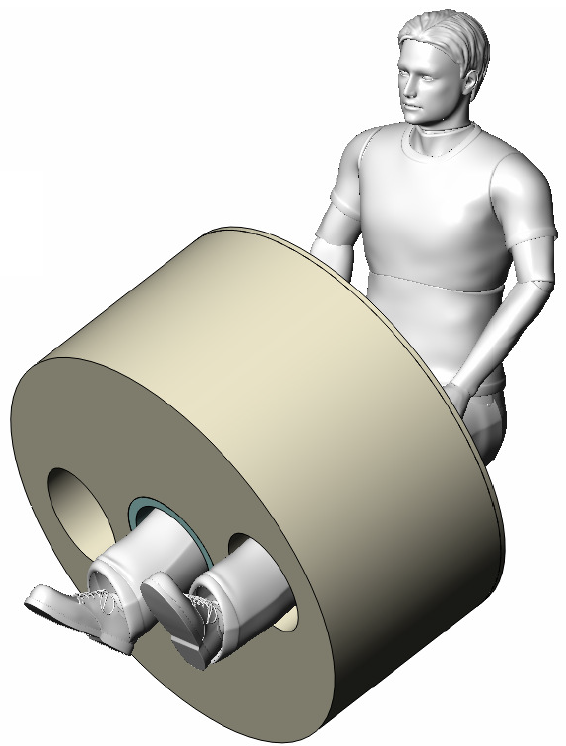
Conventional superconducting magnets use liquid helium for their cooling. Logistical complexities associated with the service of liquid helium and managing (venting) a large volume of helium gas following a quench have been among the main barriers to MRI scanners from entering point-of-care use and use in the physician’s office. To remove these barriers, the magnet was designed to be conduction cooled with a cryocooler. It requires no liquid cryogens for cooldown or normal operation; a quench results in warming of the coldmass, which merely needs to be cooled back to operating temperature in order to charge it back up to field, a process that takes 4 hours. The absence of cryogenic fluids permits the magnet to be moved and tilted while charged. To accommodate knee scanning comfortably without awkward positioning of the leg not being scanned, the magnet was constructed with three warm bores (Figure 1). The central 295 mm bore is the “actual” bore with the homogeneous field, and the truncated oval bores on either side of the central bore are placed to accommodate the leg not being scanned. The shielding coils of the central bore encompass all three bores to permit the “dummy” bores to be close to the central bore.Results
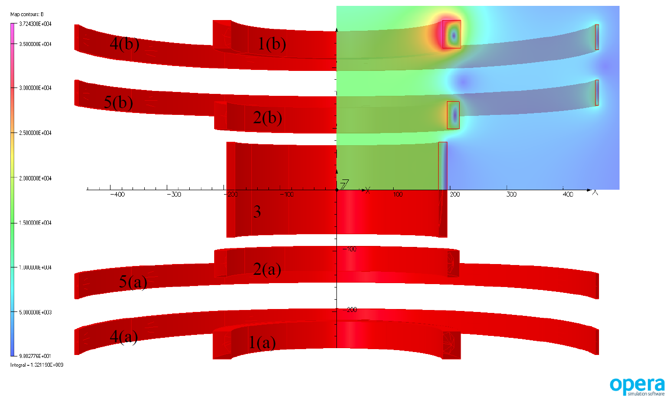
A nine coil design was optimized via an internally developed magnetic design simulation using Opera Simulation Software (Cobham Technical Services, Oxfordshire, UK), to produce a 1.5 T target field at 120.5 A current, and an oblate ellipsoidal homogeneous (5 ppm) volume with radii of a = b = 180 mm and c=160 mm, in a short 550 mm warm bore (Figure 2). The magnet was equipped with a Sumitomo SRP-082 cryocooler with a first stage cooling capacity of 40 W at 45 K and second stage capacity of 1.0 W at 4.2K. It required about four days to cool from room temperature to 3.7 K. The 5 Gauss line is about 1.70 m from isocenter on axis and about 1.50 m radially. The magnet may be ramped in 1 hour. The field exhibits no measurable drift over one day, but shows a small short term cyclical excursion of 0.4 ppm associated with the displacement of the coldhead regenerator. This was reduced to about 0.15 ppm with the installation of a small superconducting shield.Discussion
The work demonstrates the basic feasibility of this novel three-bore cryogen-free highly compact magnet design (Figure 3). The regenerator-associated field variation can likely be reduced further with more extensive shielding and/or room temperature B0 compensation. In its current state, the magnet is equipped with a pulse tube cryocooler which permits only very limited tilting, but can be cooled with a Gifford-McMahon unit that permits full tilting with somewhat higher vibration and expense.Conclusion
The cryogen-free three-bore compact superconducting magnet is practical and could form the basis of a dedicated extremity MRI scanner.Acknowledgements
Funding was provided by National Institute of Arthritis and Musculoskeletal and Skin Diseases grant R44AR065903.References
No reference found.Figures
Figure
1. Tiltable cryogen-free three-bore compact superconducting magnet concept. The
central bore is active, and one of the side bores comfortably accommodates the
knee not under examination.
Figure
2. Nine coil magnetics design.

Figure 3. The
magnet installed in a cramped laboratory.