4328
Concurrent use of 4 gradient axis enables eddy current compensation of an unshielded gradient insert coil1Radiology, University Medical Center Utrecht, Utrecht, Netherlands, 2Danish Research Centre for Magnetic Resonance, Copenhagen University Hospital Hvidovre, Denmark, 3Spinoza Center for Neuroimaging, Amsterdam, Netherlands
Synopsis
In this work, a method is presented where a whole-body gradient system corrects for eddy currents induced by an unshielded gradient insert coil. A single-axis unshielded gradient breast insert coil was positioned in a 7 tesla whole body MR system. Field cameras were used to analyze the eddy currents and validate the proposed method.
Purpose
Gradient performance is an important limiting factor for obtaining high resolution images. In particular echo planar imaging sequences can benefit from higher slew rates and gradient strengths. The introduction of actively shielded gradient coils boosted to performance of gradient systems greatly1. Since then, many effort has been put in designing high performance gradient systems, with currently the whole-body gradient system of the human connectome project being the state-of-the-art whole-body gradient system2 (300mT/m, 200T/m/s). Alternatively insert gradient systems for a single body part3 (85mT/m, 708T/m/s) can be used, which is particularly interesting for obtaining higher slew rates. To achieve the best of both worlds, both gradient insert coils and whole-body systems can be employed simultaneously4. However, actively shielded gradient insert coils are often heavy and difficult to handle in a daily setting. Therefore, these coils are not used very often. In this work we suggest to use a single-axes unshielded gradient insert coil, making the setup relatively light weight, in combination with a three-axis whole-body gradient system. The lack of shielding will, however, result in eddy currents in the different conducting parts of the magnet, which can possibly not be corrected by the insert coil itself. Here we show that the whole-body gradient system can be used simultaneously to counteract the effects of these eddy currents.Methods
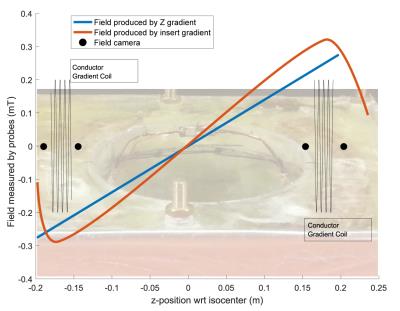
Measurements were performed with a home-built breast gradient insert coil5, in a Philips 7 tesla MR system. The scanner software was modified to enable controlling four gradient axis simultaneously, including eddy current compensation (ECC) for the self and cross terms. Four field cameras (Skope Magnetic Resonance Technologies AG, Zürich, Switzerland) were positioned in the gradient coil to analyze the eddy currents (figure 1).
Short term eddy currents
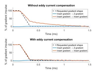
To analyze the short term eddy currents, the gradient insert coil was switched from 0.9mT/m to 0mT/m, with a ramp duration of 0.2ms. One acquisition was made without ECC, followed by an acquisition with self-term compensation using an amplitude and time constant matching the eddy current response. For both acquisitions, eddy currents were measured for 1.5ms with a temporal resolution of 1µs.
Long term eddy currents
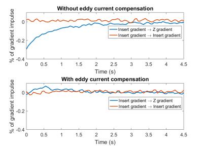
To analyze the long term eddy currents, the gradient insert coil was switched from 1.75mT/m to 0mT/m. One acquisition was made without ECC, followed by an acquisition with cross-term compensation using an amplitude and time constant matching the eddy current response. For both acquisitions, eddy currents were measured for 4.5s with a temporal resolution of 50ms.
Results
Short term eddy currents
Without ECC, almost 1ms is required to reach the desired gradient strength (figure 2, top). With ECC, with an amplitude of 60% and a time constant of 0.3ms, the desired gradient strength is reached almost instantaneously (figure 2, bottom). In both cases, practically no cross terms could be observed in this short time frame (1.5ms) of the measurements.
Long term eddy currents
When measuring the long term eddy currents, without ECC, an eddy current is measured creating a z gradient (figure 3, top), likely caused by an eddy current in the heat shield of the magnets cryostat. After enabling ECC, here a cross-term from the insert gradient to the whole-body z gradient, with an amplitude of -0.54% and a time constant of 1s, this eddy current is substantially reduced (figure 3, bottom). No long self-term eddy currents (between 0 and 4.5s) have been observed in both cases.
Discussion
A synergistic use of a gradient insert coil and a whole-body gradient system was presented. Using both systems simultaneously enables the correction of eddy currents induced by an unshielded gradient insert coil. Here, only two terms were analyzed: the self-induced eddy currents of the gradient insert coil, and cross-term eddy currents creating a whole-body z-gradient. More field cameras, with a wider spatial coverage, are required to analyze other terms, such as a x-gradient or y-gradient profiles. However, this was not feasible in this study, due to geometrical constraints only a limited number of field cameras (n=4) fitted in the coil. The rational of using a single-axis gradient insert coil comes from the fact that, when looking at EPI sequences, the highest demand can be found on a single-axis imaging gradient: the readout direction. However, the proposed method of eddy current compensation can be extended to a three axes unshielded gradient insert coil. To conclude, a whole-body gradient coil can be used for driving pre-emphasis currents to compensate eddy current effects induced by an unshielded gradient insert coil. This substantially simplifies the design of gradient insert coils.Acknowledgements
No acknowledgement found.References
1. Chapman B, Turner R, Ordidge RJ, Doyle M, Cawley M, Coxon R, Glover P, Mansfield P. Real-time movie imaging from a single cardiac cycle by NMR. Magn. Reson. Med. [Internet] 1987;5:246–254. doi: 10.1002/mrm.1910050305.
2. Setsompop K, Kimmlingen R, Eberlein E, et al. Pushing the limits of in vivo diffusion MRI for the Human Connectome Project. Neuroimage [Internet] 2013;80:220–233. doi: 10.1016/j.neuroimage.2013.05.078.
3. Tan ET, Lee S-K, Weavers PT, Graziani D, Piel JE, Shu Y, Huston J, Bernstein MA, Foo TKF. High slew-rate head-only gradient for improving distortion in echo planar imaging: Preliminary experience. J. Magn. Reson. Imaging [Internet] 2016;44:653–664. doi: 10.1002/jmri.25210.
4. Parker DL, Goodrich KC, Hadley JR, Kim S-E, Moon SM, Chronik BA, Fontius U, Schmitt F. Magnetic resonance imaging with composite (dual) gradients. Concepts Magn. Reson. Part B Magn. Reson. Eng. [Internet] 2009;35B:89–97. doi: 10.1002/cmr.b.20134.
5. van der Velden TA, van Houtum Q, Gosselink MJM, Luijten PR, Boer VO, Klomp DWJ. Characterization of a breast gradient insert coil at 7 tesla with field cameras. In: International society of magnetic resonance in medicine. ; 2016. p. 3543.
Figures


