4319
The potential of a 256-Channel receive-only Array Coil for accelerated Cardiac Imaging at 3TBernhard Gruber1,2, Arjan D. Hendriks 1, Cezar B.S. Alborahal3, Bas Brussen3, Tim Leiner1, Gustav Strijkers1, Dennis W. J. Klomp1,3, and Martijn Froeling1
1Department of Radiology, University Medical Center Utrecht, Utrecht, Netherlands, 2Institute of Biomedical Mechatronics, Johannes Kepler University, Linz, Austria, 3MR Coils B.V., Zaltbommel, Netherlands
Synopsis
High-density coil arrays can be used to accelerate MRI. Here we present the results from measurement-based extrapolations of a 256 Channel Cardiac Array Coil obtained by 16 sequential scans of a 16 Channel Array to assess acceleration performance and sensitivity constraints for 3T MRI. With element sizes of 55 mm x 33 mm, tissue load remains dominant at the 3T Larmor frequency of water, while SENSE accelerations can go up to 20-fold at low g-factors. These results motivate the design of a 256 channel cardiac array for accelerated 3T MRI.
Purpose
Todays Cardiac Magnetic Resonance Imaging (CMR) protocols can be extremely time consuming, expensive, and uncomfortable for the patient. Particularly, when including the realm of multi parametric MRI features that aid diagnosis. In order to substantially accelerate the image acquisitions, high-density receiver coil arrays1-3 may be used. Increasing the density of coils coincides with reducing the coil size, which in turn will reduce tissue loading and therefore can increase the noise figure of the MRI system. But, as long as tissue load remains dominant, the density of the coils can be increased without substantially adding noise4. In this study, we have investigated the use of high-density coil arrays for accelerated5-7 cardiac MRI while assuring tissue load dominance. In process of building the receiver chain, 16 sequential measurements with a repositioned high density 16-channel receiver array are performed to obtain coil sensitivity and noise correlations in a healthy volunteer to investigate the possible acceleration performance of a 256 Channel Cardiac Coil Array at 3 Tesla.Methods
A 16-element (4 x 4) coil array was designed (MR Coils BV, Zaltbommel, the Netherlands) with loop sizes of 55 mm in length and 33 mm in width (Fig. 1). Individual Q-factor measurements were performed using an S12 measurement with two pick-up probes weakly coupled (<<-30dB) to the loop. Unloaded and loaded Q values were measured at different locations of the body. Measurements were performed on a Philips 3T wide bore system with digital receivers (Philips, Best, the Netherlands). Coil sensitivity maps were recorded using the standard Philips reference scan (resolution = 6 mm isotropic, two stacks: one obtained with 16 channels as receiver and one with the bodycoil as receiver). A separate noise scan was obtained by switching off the RF transmit power. To simulate the expected improvement in acceleration of the 256 Channel Cardiac Array, the measurements were repeated 16 times, each time with the array positioned at another location around the chest of the patient. The 16 data sets of the array were spatially registered and merged into a 256 by 256 array filling out only the diagonal 16 times 16 elements. The remaining part of the array is set to zero. G-factor maps are calculated with SENSE accelerations in FH and LR using the aligned sensitivity maps and the noise matrix.Results
The unloaded Q-factor was measured to be 603, while the loaded Q-factor ranged from 53-98. The noise correlation matrix is shown in Fig. 2. No noise correlation could be obtained for the cross terms outside the 16 element wide diagonal of the 256x256 matrix, due to absence of sufficient receiver chains. Note the low noise correlation within the 16-element wide diagonal. When merging the 16 images obtained with the 16 times 16 elements, good penetration of the reception is obtained throughout the body (Fig. 3). SENSE acceleration can go up to 5 in one dimension, or 4x5 in 2D acceleration (Fig. 4).Discussion
The results show that a 256 Channel Cardiac Coil allows acceleration factors of up to 20 when accepting a g-factor of less than 2. It should be noted that the results are obtained with only 16 elements physically present during each scan. Consequently, potential couplings in a full 256 Channel array have not been incorporated. However, it is expected that these potential couplings will not effect our results substantially as the elements within the 16 Channel array also show very low coupling.Conclusion
We have demonstrated that the coil element that can be used for a high-density 256 Channel Cardiac Array remains in tissue load dominance (6 to 11-fold). Moreover, coil couplings within the 4 x 4 array can be very low. When neglecting potential noise cross correlation within the remaining elements of the 256 Channel Array Coil, image acceleration of over 20 can be obtained with SENSE encoding. Consequently, these calculations demonstrate the motivation to build the 256 Channel Cardiac Array Coil to facilitate a boost in acceleration of the multi parametric cardiac MRI.Acknowledgements
No acknowledgement found.References
1. Roemer P.B. et al., MRM 16 (1990): 192-225. 2. Schmitt M. et al., MRM 59 (2008): 1431-1439. 3. Etzel R. et al., ISMRM 2015:1780. 4. Schuppert M. et al., ISMRM 2015:1779. 5. Sodickson D.K. et al., MRM 38 (1997): 591–603. 6. Pruessmann K. P. et al., MRM 42 (1999): 952–962. 7. Griswold M.A. et al., MRM 47 (2002): 1202–1210.Figures
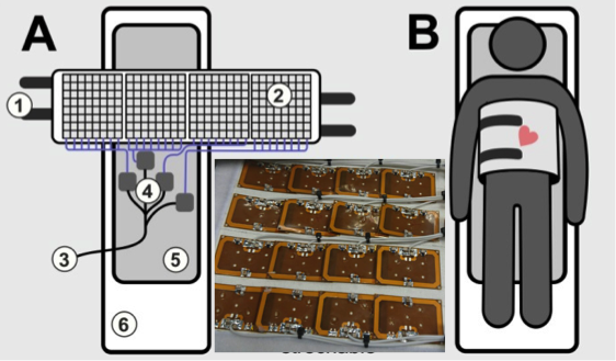
Fig. 1 - 4 x 4 high density coil array to simulate the performance
of the design of a 256 Channel Cardiac Array Coil as indicated in A and B,
incorporating cable traps, preamps and digital receivers (4), fixation straps
(1), digital interfacing (3), patient comfort (5) and the MRI table (6).
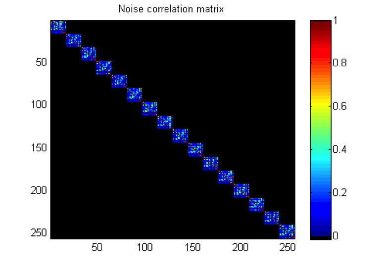
Fig. 2 - Noise Correlation between the 16 elements at the 16
different locations on the body. Note the low coupling between the elements,
and obviously the absence in the outer parts of the matrix.
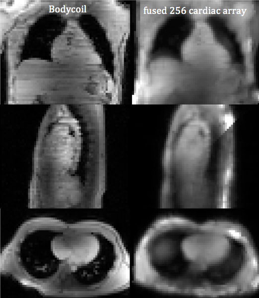
Fig. 3 - Combined MRI from the 256 locations of the receiver
coils (right) as compared to the Bodycoil (left). Note the full penetration
depth of the array of small element receivers.
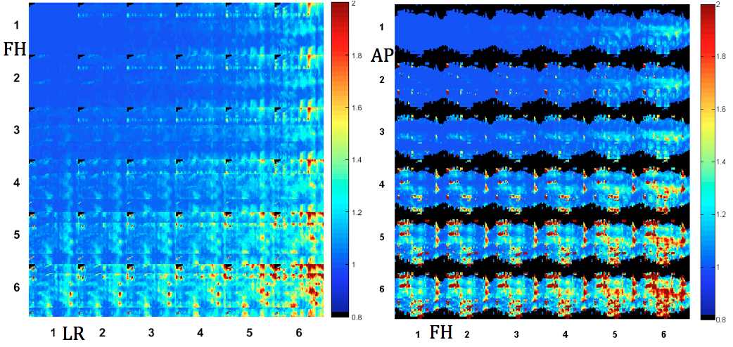
Fig. 4 - G-factor maps of two orthogonal slices from SENSE
acceleration in AP, LR and FH directions. Note that accelerations of 4 x 5 (FHxLR)
or 3 x 5 (APxFH) can be obtained while remaining a low g-factor.