4315
An open 8-channel pTx coil for 7-Tesla MRI of the knee and ankle joints at multiple postures1University of Queensland, St Lucia, Australia, 2Siemens Ltd, Brisbane, Australia, 3American University of the Middle East, Kuwait
Synopsis
This paper presents the initial in vivo imaging results from a new open 8-channel pTx transceive array designed and constructed for 7 T MRI of the knee and ankle joints. The open design of the coil provides easy access and conformable accommodation of both joints, while enabling the joints to be imaged at multiple angles for enhanced pathological assessment. With individualized RF shims, the array provided full coverages and uniform excitations for both joints, demonstrated with high quality 2D TSE and 3D GRE (DESS and MEDIC) images.
PURPOSE:
MRI of musculoskeletal (MSK) systems can potentially benefit from ultrahigh-field (UHF) systems owing to intrinsically higher SNR and thus enhanced spatial resolution. Parallel transmit (pTx) is a prevalent technique to address inhomogeneous excitation, local SAR restriction, and limited peak RF powers at UHF 1. At present, however, commercial RF coils for MSK imaging at 7 T are limited to the knee and typically do not have pTx capability. To improve transmission performance, there have been several important custom knee pTx coil developments, including a 2-channel transmit coil combined with the QED receive array 2, and a 4-channel proton birdcage sodium transceiver array for multi-nuclei imaging 3. A multi-purpose 16-channel RF array with true pTx capability was also presented with gradient-echo (GRE) imaging of the knee 4. For ankle imaging at 7 T, there is an absence of a dedicated commercial coil. Besides a custom 8-channel pTx ankle coil 5, recent studies resorted to adapting commercial head and knee coils 2, 6-9. For 7-T MRI of the knee and ankle joints, we proposed an open design pTx coil array 10. This paper presents the initial in vivo imaging results from the newly constructed array.METHODS:
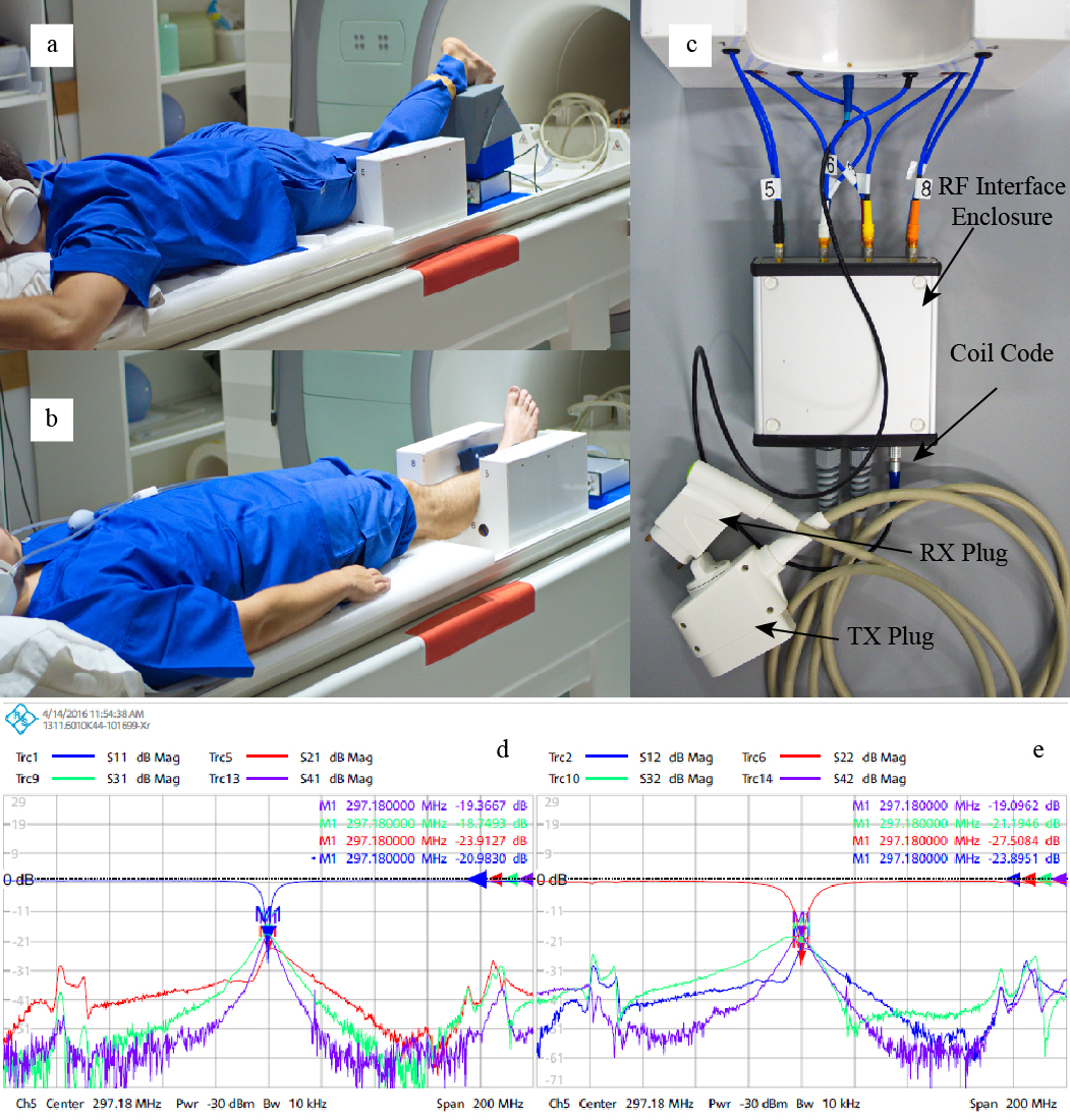
Figs. 1a - 1d illustrate the U-shaped open pTx coil and its placement relative to knee and ankle joints in 7-T imaging applications. The 8 loop elements were arranged in two cylindrical rows (diameter 180 mm) to maximize the field of view (FOV) in the head-foot direction (length 200 mm). An RF shield was placed inside the coil housing 30 mm radially away from the elements. The element rungs were made of multilayered stripline-like conductors/blades 11, which were oriented angularly to improve B1 penetration and inter-element decoupling 12. Small counter-wound inductive decoupling loops 13 were used to further decouple neighboring elements. Detailed coil geometry was optimized with numerical simulations using Sim4Life by ZMT 14 combined with two anatomically accurate voxel models (Duke and Ella 15). The electric fields of 1 V harmonic sources were extracted to calculate virtual observation points (VOP) using vendor supplied software, facilitating efficient in vivo estimation of worst-case local SAR. Healthy male volunteers were imaged for the knee (N=2, 56 and 52 years, 91 and 78kg) and ankle joints (N=2, 52 and 25 years, 78 and 83kg) using a 7-T whole-body research scanner (Siemens Healthcare, Erlangen, Germany), after obtaining written consent. Both turbo spin echo TSE (with PD, PD + fat saturation, T1 and T2 weightings) and GRE sequences (3D water-excited Dual-Echo Steady-State we-DESS and 3D Multi-Echo Data Image Combination MEDIC) were tested, as detailed below.RESULTS:
The measured S-parameters for the new coil showed a good agreement with the simulated results, with S11 values of all channels better than -23 dB, and S1X -19 dB (Figs.1d and 1e).
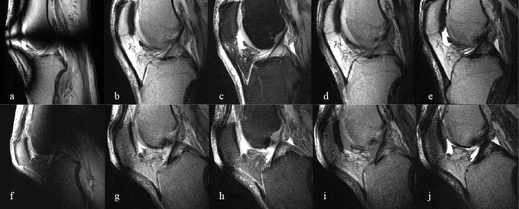
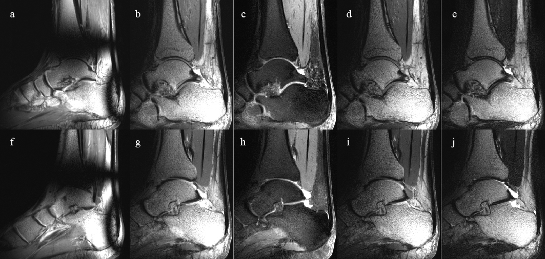
Fig.2 shows representative sagittal TSE images (FOV = 160 mm, resolution = 0.3 × 0.3 × 2.5 mm3, TA < 4 min, #slices = 25) of the left knee joints in extended and flexed postures, both of which provide excellent visualization of the anterior cruciate ligament (ACL). Similarly, Fig.3 illustrates the representative TSE images of the ankle joint in neutral position, providing visualization of the Achilles tendon and the interosseous talocalcaneal ligament. The circularly-polarized mode used at least twice as much peak RF power as the shim modes, and produced strong artifacts. With subject-specific shims, the examination was performed well under the regulatory SAR limits. The highest 10sec-averaged SAR was observed for the knee T1w TSE sequence at 9.71 W/kg.
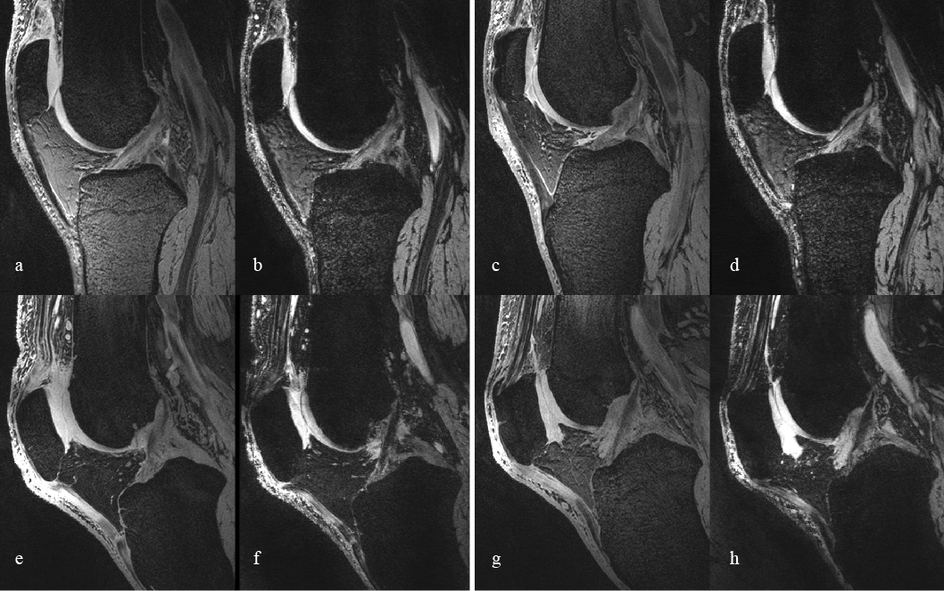
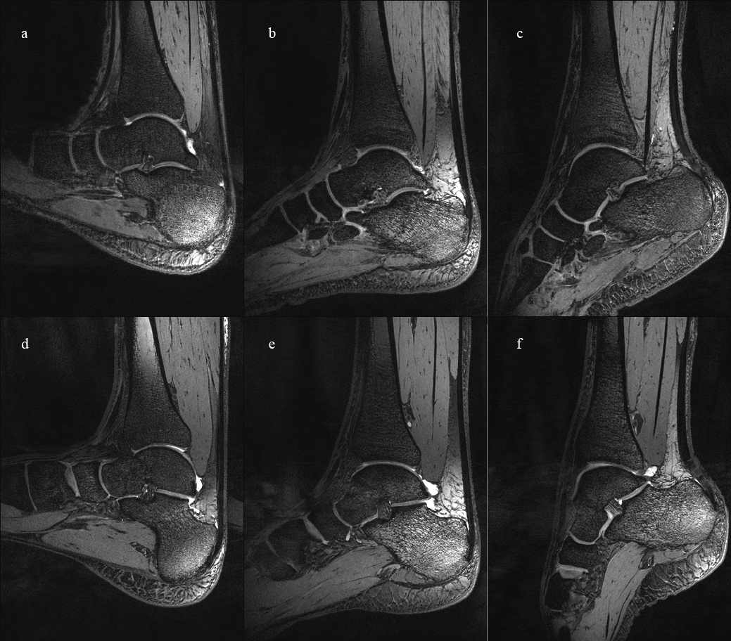
Fig.4 displays representative sagittal 3D we-DESS and MEDIC images of the knee joints of the two volunteers. Both sequences had a FOV = 210 mm, resolution = 0.47 mm isotropic, and TA = 5:30 min. Fig.5 displays the sagittal views of the ankle joints acquired using the 3D we-DESS acquired at maximal dorsiflexion, neutral, and maximal plantarflexion postures.
DISCUSSION:
High quality 2D (TSE) and 3D (DESS, MEDIC) images of the knee and ankle joints at multiple joint postures were acquired from a new 8-channel pTx array (dynamic imaging is also possible 16). Combined with successful subject-specific shims, the coil array provided full joint coverage and excellent image homogeneity over the region of interest for all sequences. The 3D GRE sequences provided excellent contrast between the joint cartilage, surrounding fluids and the cortical bones. Some incomplete water excitation was observed with DESS and may be further improved with better B0 shimming and frequency adjustments.CONCLUSION:
The initial high-quality knee and ankle images at various joint postures were presented for the new 7-T open pTx RF coil array.Acknowledgements
We acknowledge ZMT Zurich MedTech AG (www.zurichmedtech.com) for providing the electromagnetic simulation software package SIM4LIFE. All correspondence should be made to J. Jin (jinjin@itee.uq.edu.au).References
[1] H. Homann, I. Graesslin, H. Eggers, et al., "Local SAR management by RF Shimming: a simulation study with multiple human body models," Magnetic Resonance Materials in Physics, Biology and Medicine, vol. 25, pp. 193-204, 2012/06/01 2012.
[2] P. E. Z. Larson, M. Han, R. Krug, et al., "Ultrashort echo time and zero echo time MRI at 7T," Magnetic Resonance Materials in Physics, Biology and Medicine, vol. 29, pp. 359-370, 2016.
[3] R. Brown, G. Madelin, R. Lattanzi, et al., "Design of a nested eight-channel sodium and four-channel proton coil for 7T knee imaging," Magnetic Resonance in Medicine, vol. 70, pp. 259-268, 2013. [4] B. Wu, X. Zhang, C. Wang, et al., "Flexible Transceiver Array for Ultrahigh Field Human MR Imaging," Magnetic resonance in medicine : official journal of the Society of Magnetic Resonance in Medicine / Society of Magnetic Resonance in Medicine, vol. 68, pp. 1332-1338, 01/13 2012.
[5] S. Orzada, A. K. Bitz, L. C. Schäfer, et al., "Open design eight-channel transmit/receive coil for high-resolution and real-time ankle imaging at 7 T," Medical Physics, vol. 38, pp. 1162-1167, 2011.
[6] J. M. Theysohn, O. Kraff, S. Maderwald, et al., "MRI of the ankle joint in healthy non-athletes and in marathon runners: image quality issues at 7.0 T compared to 1.5 T," Skeletal Radiology, vol. 42, pp. 261-267, 2013// 2013.
[7] V. Juras, G. Welsch, P. Bär, et al., "Comparison of 3 T and 7 T MRI clinical sequences for ankle imaging," European Journal of Radiology, vol. 81, pp. 1846-1850, 8// 2012.
[8] V. Juras, S. Zbyn, C. Pressl, et al., "Regional variations of T2* in healthy and pathologic achilles tendon in vivo at 7 Tesla: Preliminary results," Magnetic Resonance in Medicine, vol. 68, pp. 1607-1613, 2012.
[9] X. Deligianni, P. Bär, K. Scheffler, et al., "High-resolution Fourier-encoded sub-millisecond echo time musculoskeletal imaging at 3 Tesla and 7 Tesla," Magnetic Resonance in Medicine, vol. 70, pp. 1434-1439, 2013.
[10] B. Henin, E. Weber, and S. Crozier, "A Novel 8-channel transceive open knee coil for dynamic musculoskeletal imaging at 7 Tesla," in Proceedings of the 24th Annual Meeting of ISMRM, Singapore, 2016.
[11] E. Weber, B. K. Li, F. Liu, et al., "A ultra high field multi-element transceive volume array for small animal MRI," in Engineering in Medicine and Biology Society, 2008. EMBS 2008. 30th Annual International Conference of the IEEE, 2008, pp. 2039-2042.
[12] B. K. Li, F. Liu, E. Weber, et al., "Hybrid numerical techniques for the modelling of radiofrequency coils in MRI," NMR in Biomedicine, vol. 22, pp. 937-951, 2009.
[13] S. Crozier, B. K. Li, F. Liu, et al., "MRI coil design," WO2009124340 A1, 2009.
[14] SIM4LIFE by ZMT. Available: www.zurichmedtech.com
[15] C. Andreas, K. Wolfgang, G. H. Eckhart, et al., "The Virtual Family—development of surface-based anatomical models of two adults and two children for dosimetric simulations," Physics in Medicine and Biology, vol. 55, p. N23, 2010.
[16] A. Destruel, K. O'Brien, J. Jin, et al., "Dynamic imaging of the knee and ankle joints at 7T," in Proceedings of the 25th Annual Meeting of ISMRM, Honolulu, HI, USA, 2017, p. 5489.
Figures