4303
Experimental realization of a novel dual-nuclei coil for small animal imaging at 7 Tesla based on periodic structures of metal strips1Department of Nanophotonics and Metamaterials, ITMO University, Saint-Petersburg, Russian Federation, 2DRF/I2BM/Neurospin/UNIRS, CEA-Saclay, Paris, France, 3CNRS, Institut Fresnel, Aix-Marseille Université, Marseille Cedex, France
Synopsis
In order to acquire image or MR spectra at two different nuclei dual-frequency RF-coils are used. Conventional approaches of tuning and matching a coils at several frequencies employ expensive non-magnetic capacitors which also introduce dissipative losses. We propose an alternative method of tuning and matching the coils at multiple desired frequencies. This method is based on resonant excitation of hybridized eigenmodes of periodic structures of metal strips. The proposed method allows one to build cheap coil with reduced dissipative losses resonating on two different frequencies simultaneously.
Purpose
Dual-frequency RF coils are widely used in MRI and MRS to acquire images or MR spectra at two different nuclei. There are several conventional approaches for tuning and matching RF coils at multiple frequencies. One of them includes a single loop tuned to and matched at two frequencies by means of a lumped elements circuitry [1,2]. The other method employs two separate coupled loops [3]. Both methods involve expensive tunable non-magnetic capacitors which disadvantageously introduce dissipative losses. In this work we propose and experimentally study an alternative method of tuning and matching of dual-frequency coils for small-animal MRI simultaneously at two nuclei: fluorine 19F and proton 1H having Larmor frequencies of 282.6 and 300.1 MHz respectively at 7.05 Tesla. Since the proposed coil does not require any lumped elements, this design is cheap and has reduced losses.
Methods
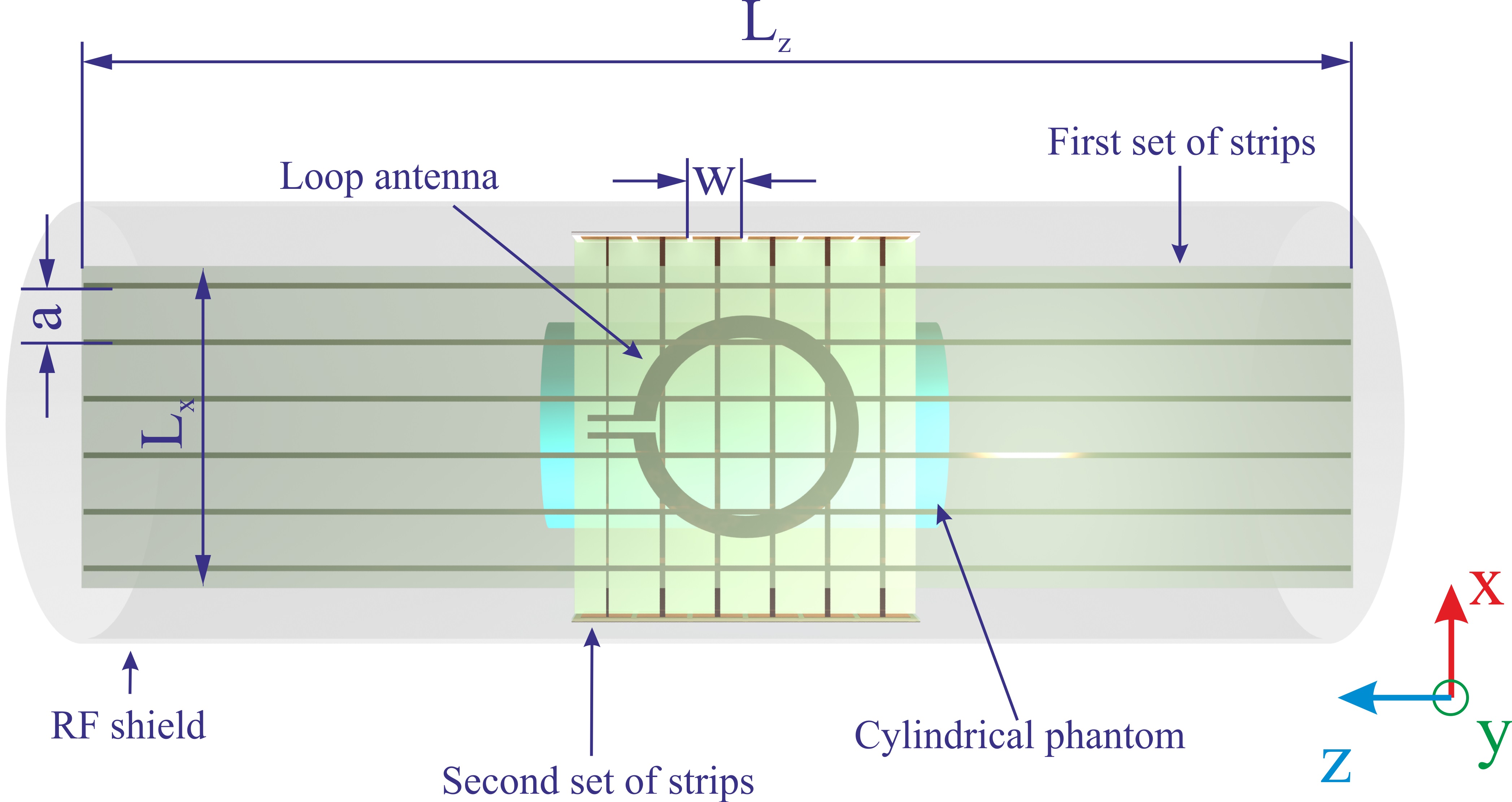
The proposed coil contained two mutually orthogonal sets of metal strips each one resonating at one of the desired frequencies. Both sets of strips were implemented using printed circuit boards (PCBs) on a 0.508 mm-thick substrate (Rogers 4003, ε = 3.38, tan δ = 0.002). We used identical strips with the period of 10 mm and the width of 1 mm. In each periodic set of resonant strips a number of hybridized eigenmodes can be excited using a small magnetic loop [4,5].
We performed numerical simulation of the coil with CST Microwave Studio. In the coil the first set of six strips in parallel to B0 axis had the length of 451 mm, which is close to the half-wavelength. The loop excites the lowest-frequency eigenmode of the first set of strips at 282.6 MHz. Second set of resonant strips was placed orthogonally to the first one 1 mm above. Since the available space was limited by the bore diameter, the second set was composed of 72 mm strips. These short strips were made resonant at 300.1 MHz by loading on distributed capacities [6] implemented as square printed patches (Rogers 4003, 0.508 mm). The structure (Figure 1) was fed by a printed circular loop (radius 20 mm). The coil was simulated in the presence of the 90-mm RF shield of a preclinical MRI machine and the homogeneous cylindrical phantom (ε = 39, σ = 0.25 S/m) of the diameter 40 mm mimicking properties of a small animal.
The proposed coil was manufactured and investigated experimentally. The coil was constructed of five separate PCBs (two supporting strips, two supporting patches and one supporting the loop). Each strip of the second periodic set was soldered to a patch from both ends. The homogeneous cylindrical phantom was made of a plastic container with the mixture of 60% 2-2-trifluorethanol and 40% water. In order to evaluate matching and tuning properties of the coil its parameter S11 was measured by means of the VNA Anritsu MS2027C. The measurements were performed in the environment of the preclinical MR scanner Bruker PharmaScan 7 Tesla. It was shown that one can match and tune the coil to 282.6 and 300.1 MHz simultaneously by varying the mutual position of the two sets of strips and the loop without any lumped elements.
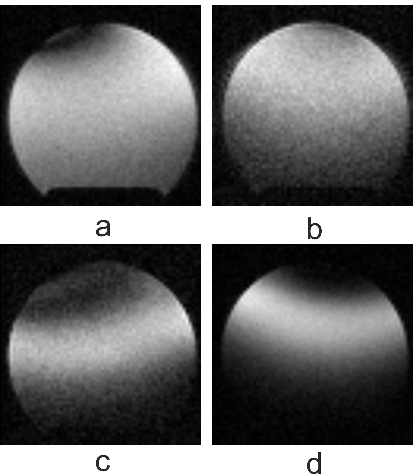
On the next step, we acquired MR images of the phantom at the two Larmor frequencies. The scans were made using the manufactured antenna on the same preclinical system (both gradient T1 FLASH (TR/TE=2000/2.4 ms, 0.7x0.7x0.7 mm3), and spin echo T2 TurboRARE (TR/TE=2000/4.6 ms, 0.7x0.7x0.7 mm3) sequences).
Results
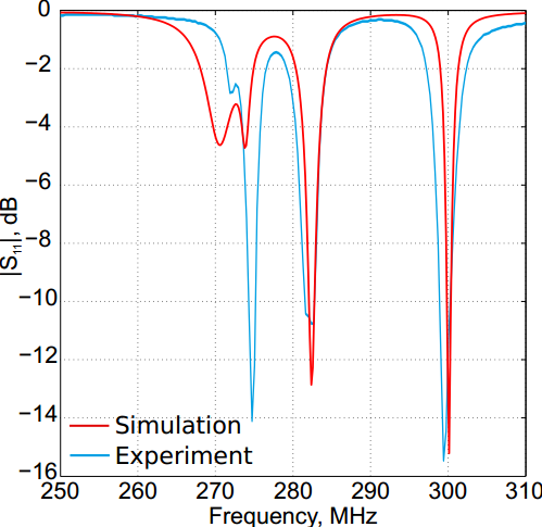
Figure 2 shows both numerical and experimental S11 curves. The red curve corresponds to numerical simulation, while the blue one represents the measured S11. The curves demonstrate good agreement between the theory and the experiment. As one can see from Figure 2, it was possible to reach the level of S11 below -10 dB at both the desired Larmor frequencies simultaneously.
Figure 3 shows the reconstructed images of the central axial plane of the phantom, acquired both for fluorine (a,c) and proton (b,d). Figures 3 (a,b) show the MR images obtained with the gradient echo sequence, while Figures 3 (c,d) exhibit the images acquired by the spin echo sequence. The obtained levels of SNR were 18.8 and 34.8 for proton, 32.4 and 22.5 for fluorine (by gradient and spin echo respectively).
Conclusions
The new design of a dual-frequency RF-coil for 19F and 1H preclinical imaging based on the resonant excitation of hybridized eigenmodes of periodic sets of metal strips was realized and investigated experimentally. The coil was tuned and matched without expensive non-magnetic capacitors simultaneously at both frequencies which was shown by simulation and measurements.Acknowledgements
This work was supported by the Russian Science Foundation (Project No. 15-19-20054).References
1. Doty at al. 'Radio frequency coil technology for small-animal MRI' NMR in Biomedicine, 20, pp. 304325, 2007
2. Hu et al. 'A generalized strategy for designing 19 F/1H dual-frequency MRI coil for small animal imaging at 4.7 Tesla' J Magn Res Imag, 34, pp. 245252, 2011
3. Alecci et al. 'Practical design of a 4 Tesla double-tuned RF surface coil for interleaved 1H and 23 Na MRI of rat brain' J Magn Res,181(2), pp. 203-211, 2006,
4. Slobozhanyuk et al. 'Enhancement of Magnetic Resonance Imaging with Metasurfaces' Adv Mater, pp. 18321838, 2016
5. Jouvaud et al. 'Volume coil based on hybridized resonators for magnetic resonance imaging' Appl Phys Lett, 108, p. 023503, 2016
6. Glybovski et al. 'Capacitively-loaded metasurfaces and their application in magnetic resonance imaging' pros of RADIO, 2015
Figures