4300
Comparison of different RF coil designs for short T2* samples1Dept. of Radiology, Medical Physics, Medical Center – University of Freiburg, Freiburg, Germany
Synopsis
MRI of short T2* samples is possible with ultra-short echo time sequences (UTE) which can be further improved using dedicated RF coils optimized for short-TE imaging. This work compares the performance of a commercial wrist coil with two custom-built coils (birdcage, solenoid) for UTE imaging of a mummy hand (low water content, short T2*), and an in-vivo measurement of a human hand in which short-T2* tissues such as tendons are highlighted by UTE image subtraction.
Introduction
MRI provides an exceptional soft tissue contrast for long T2* times, but is limited in the distinction of extremely short T2* times such as in bones, tendons and dehydrated tissues. Recent publications present methods of acquiring short T2* signals with adapted sequences such as ultrashort echo time (UTE)1, single point imaging (SPI) and pointwise encoding time reduction with radial acquisition (PETRA)2. However, RF coils and hardware elements are equally crucial for short T2* measurements. Conventional RF coils have long coil ring down times and an abundance of hydrogen in the housing material that contribute to the MR signal which limits the quality of short T2* acquisitions3. Therefore, the need for special designed RF coils for ultrashort echo time measurements arises. In this work, a comparison of a commercial circularly polarized (CP) wrist coil with different custom coils in terms of image quality and SNR is presented.Material and Methods
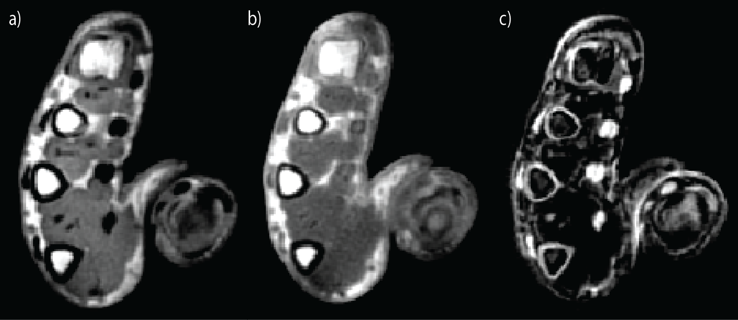
An ancient Egyptian mummy hand (ca. 1500-1100 BC, former collection of Musée D’Orbe, Suisse) was used as a short T2* sample. All experiments were performed on a 3 T clinical MR system (Prisma, Siemens Healthcare, Erlangen). A 3D UTE sequence was applied with following parameters: TE=70 µs, TR=3 ms, FoV=190x190x190 mm3, BW=1680Hz/Pixel, α=16° and 100000 radial spokes with 192 points per spoke. Images were reconstructed onto a 192x192x192 matrix using Kaiser-Bessel gridding. A quadrature Tx/Rx low pass birdcage coil with a diameter of 9 cm, 8 legs, Q factor of 150 and a ring down time of 0.4 µs was designed. A solenoid Tx/Rx RF coil with a diameter of 9 cm, Q factor of 650 and a ring down time of 1.6 µs was also constructed. For comparison, data were acquired with two custom-made coils and the commercial CP Wrist Array Coil (Siemens). For high SNR 4 averages were acquired with a total acquisition time of 33 minutes. A second data set were acquired in an in-vivo experiment with the hand of a volunteer comparing the birdcage coil and the wrist coil with the same UTE sequence and following altered parameters: 1 average, 50000 spokes, TR=6 ms and α=8° corresponding to an acquisition time of 5 minutes. To highlight short T2* components, a subtraction image is generated by two MR images with different TEs4: TE1=2000 µs and TE2=70 µs for the birdcage coil and TE1=2000 µs and TE2=150 µs for the wrist coil. To correct for motion, the image acquired with the longer echo time was co-registered to the image with shorter echo time.Results
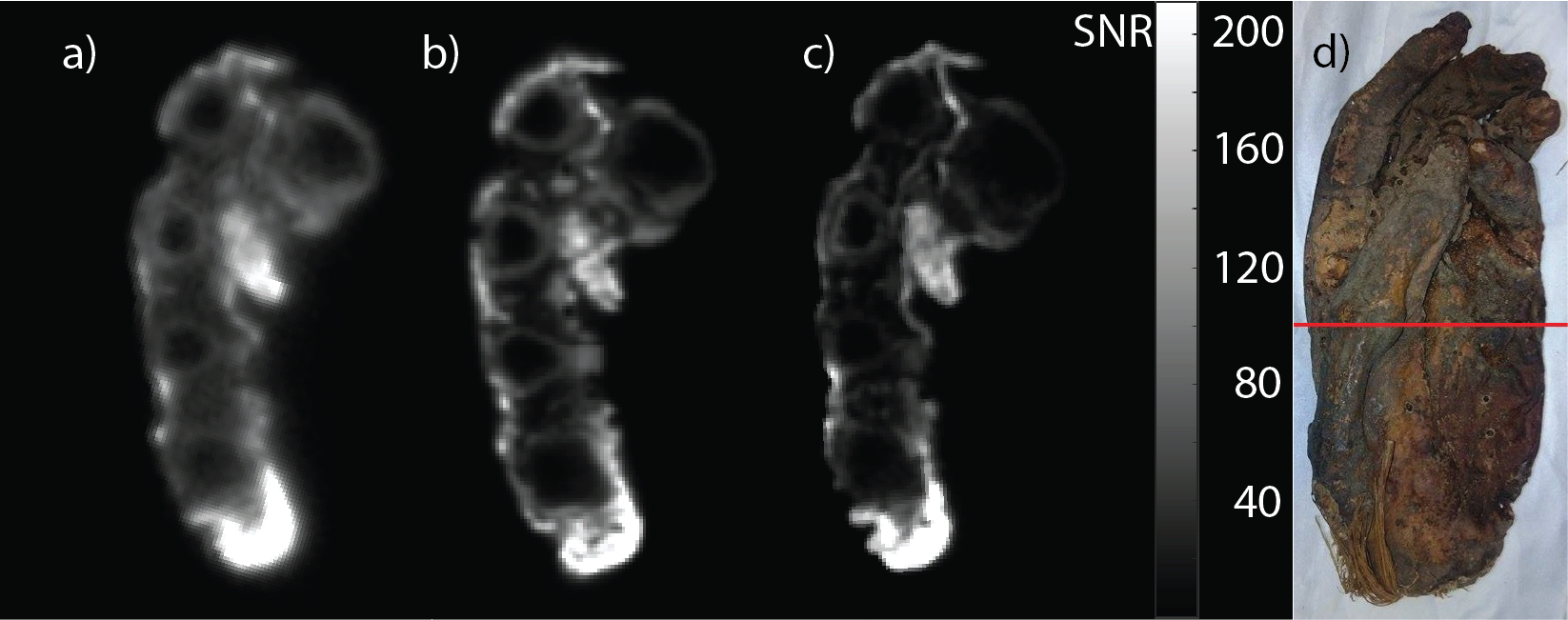
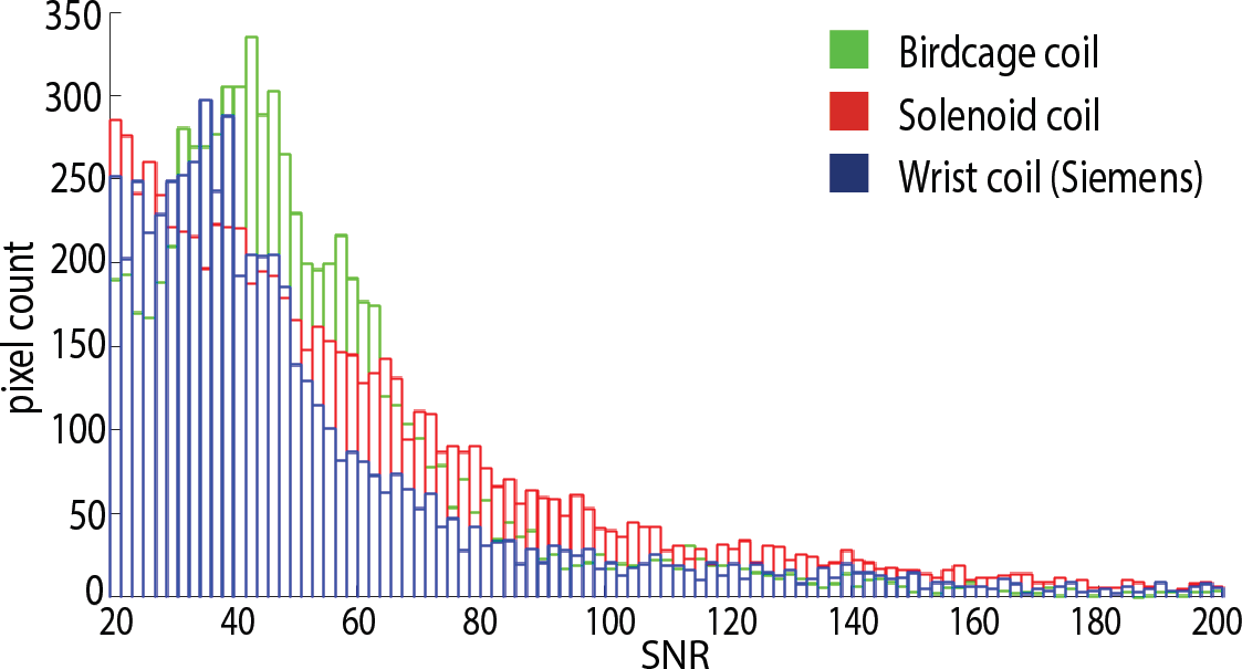
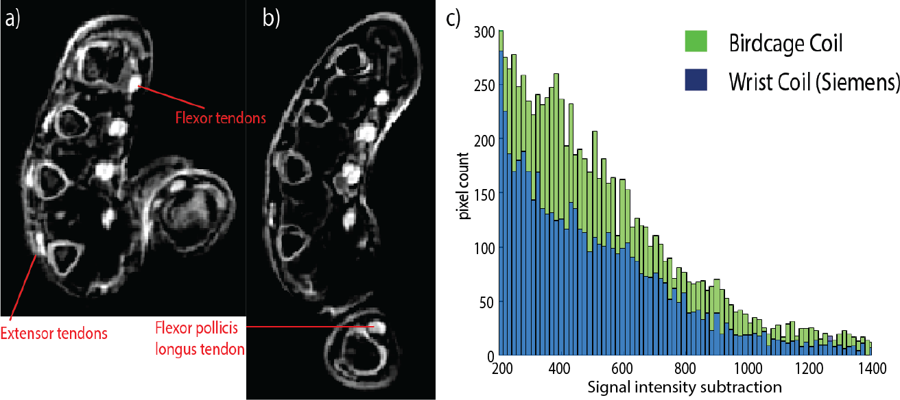
UTE images of the mummy hand were successfully acquired with all coils (Fig.1). The birdcage coil images showed the highest SNR, but these images have an apparent overall blurring which hampers the differentiation of anatomical details. The solenoid coil offers a lower SNR but retains more detail information, and the wrist coil showed fine structural details, but provided the lowest SNR and contrast. Figure 2 shows a comparison of the SNR histograms of all three coils: for this setup, the birdcage coil shows an SNR maximum in the range of 20-60. Figure 3 shows the results of the in-vivo measurements at two different TE. The subtraction image highlights tissue with short T2* such as tendons and, to a lesser extent, of bones and periosteum. Figure 4 presents a direct comparison between the subtraction images, acquired at different echo times using the birdcage and the wrist coil. Both images show a high intensity at the location of the tendons and cortical bones. However, the birdcage coil displays more signal intensity for remaining anatomical structures compared to the wrist coil. This is illustrated in the subtraction histogram of both coils where the birdcage coil has a higher signal intensity subtraction count for the entire range of 200 to 1400.Discussion and Conclusion
All MR images of the mummy hand show fine anatomical details. Birdcage and solenoid coil have higher pixel count in the range of 20-100 SNR compared to wrist coil due to higher filling factor (9 cm diameter of custom coils compared to rectangular 7x14 cm shape of wrist coil). Since TE of wrist coil is limited to 150 µs, the signal from tissue components with shorter T2* will be lost. The quadrature driven birdcage coil might provide more sensitivity compared to solenoid coil, where sensitivity drops with increasing distance from feeding port. Ring down times and switching times of commercial coils increase the dead time, therefore custom designed RF coils and circuitry are favored.
Acknowledgements
Grant support from the Deutsche Forschungsgemeinschaft (DFG) under grant numbers BO 3025/8-1 and UL 1187/6-1 is gratefully acknowledged.References
1. Tyler J, et al. Magnetic resonance imaging with ultrashort TE (UTE) PULSE sequences: technical considerations." Journal of Magnetic Resonance Imaging 25.2 (2007): 279-289.
2. Özen AC, et al. Comparison of ultrashort echo time sequences for MRI of an ancient mummified human hand. Magnetic resonance in medicine 75.2 (2016): 701-708.
3. Horch A, et al. RF coil considerations for short-T2 MRI. Magnetic resonance in medicine 64.6 (2010): 1652-1657.
4. Du J, et al. Short T2 Contrast with Three-Dimensional Ultrashort Echo Time Imaging. Magnetic resonance imaging 29.4 (2011): 470–482.
Figures