4296
RF Pulse Design - based Parallel Transmit Array Design1Biomedical Engineering, Vanderbilt University, Nashville, TN, United States, 2Institue of Imaging Science, Vanderbilt University, Nashville, TN, United States
Synopsis
A novel parallel transmit array design method is proposed that integrates both Maxwell and Bloch equations. The method is demonstrated to be better than traditional coils in dynamic multiband shimming and reduced field-of-view imaging scenarios to achieve better excitation accuracy and lower RF energy deposition, as well as robustness across multiple subjects and excitation schemes.
Purpose
Parallel transmission (pTx) using a multi-channel transmit array is an important technique that can be used to achieve spatially homogeneous excitation in high field MRI. It can also enable efficient reduced FOV (rFOV) excitations for high resolution imaging with short acquisition times (1). The major limitation of current pTx array design methods is their little involvement of RF pulse design principles. This results in designs that are mainly focused on the performance of individual channels instead of the synergistic interactions of all channels, thus the performance of such arrays are limited in maximizing excitation accuracy while minimizing SAR (2). In this study, a new transmit array design method that integrates the pTx pulse design principle is proposed and compared to existing coil design methods.Theory
The proposed pTx array design method is based on array-compressed parallel transmit (AcpTx) pulse design (3). Similar to the AcpTx framework that allows an M-coil array to be combined and connected to N-channels power amplifiers (M>N), the proposed coil array design method starts with electromagnetic fields from M coil elements, and then prunes and combines them (4) down to N-channels (M>>N). This study compares existing and proposed array design methods for two applications: 1. Dynamic multiband shimming in the human brain, and 2. rFOV imaging of the human occipital lobe.Methods
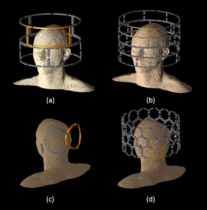
To study the effect of using a population of human models, the “Duke” human model from the virtual family (5) was used and scaled with an additional 5% ratio in X, Y, Z and all three directions respectively, creating a population of 5 subjects. They were simulated in XFDTD (Remcom Inc, State College, PA, USA) with different coil arrangements (Figure 1) to obtain EM fields at 7T --- For multiband imaging of the human brain: Case 1, Fig 1(a), a standard 2-row-by-4-coil-per-row 8 channel array; Case 2, Fig 1(b), a proposed 4-row-by-8-coil-per-row 32 channel array to be combined to 8 channels; For reduced FOV imaging of human occipital lobe: Case 3, Fig 1(c) a standard 2 channel array; and Case 4, Fig 1(d), a proposed 3-row-by-16-coil-per-row 48 channel array to be pruned to 8 loops and combined to 2 channels. For the multiband shimming cases 1 and 2, SAR virtual observation points (VOPs) were calculated (6) and used for regularization to design SAR-efficient array. The proposed coil combination method was also compared to using quadrature coil combination. For the occipital imaging cases 3 and 4, B0 fields were calculated (7) for each human model and shimmed in the target excitation region with second order shim gradients. Spiral-in 3D SPINS RF excitation k-space trajectories were designed (8). An MP-RAGE sequence with 2.6s inversion TR, 7.1ms FLASH TR, 6.5° flip angle, a 256 × 256 × 192 matrix in sagittal acquisition (9) was used to calculate maximum local 10-gram averaged SAR (10). For fair comparison, the coil size in case 3 was determined by comparing several loop sizes for the maximum rFOV excitation accuracy and the minimum SAR.Results
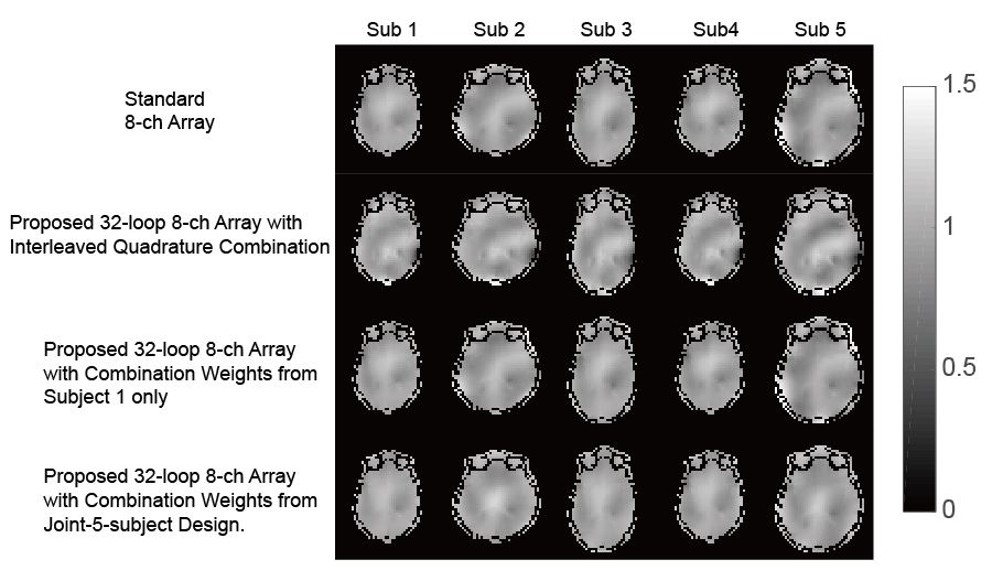
Figure 2 shows the excitation of the multiband shimming arrays. The coil pruning and combination strategy with joint-5-subject design showed better excitation accuracy than the standard 8-ch array and the quadrature combination method.
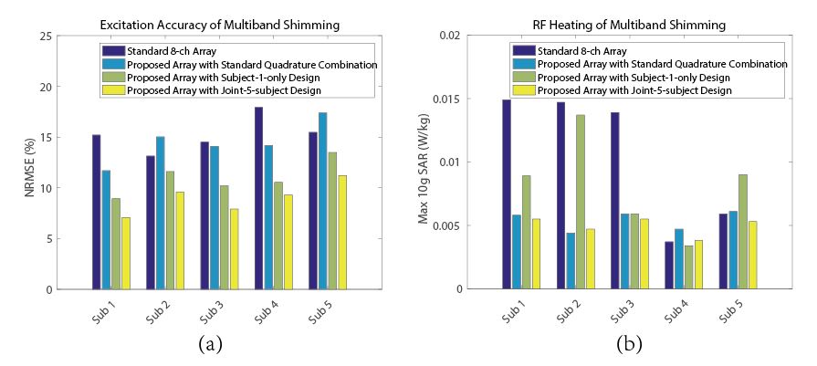
Figure 3 summarize the transmit NRMSE and the maximum 10g SAR of the multiband excitation pulses in the brain. The coil combination weights and pruning strategy obtained from the joint-5-subject design showed the best overall performance.
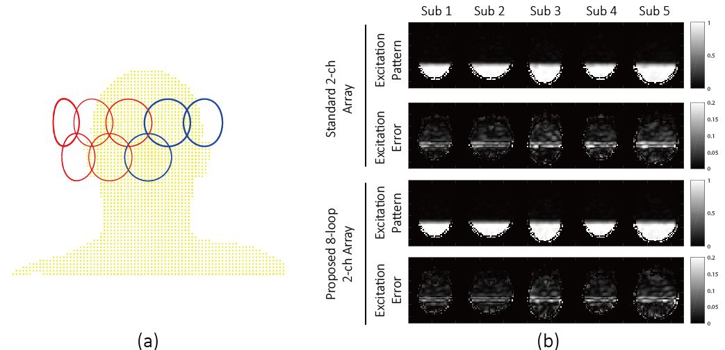
Figure 4 shows (a) the coil element arrangement of the proposed 8-loop-2-channel array, designed by jointly considering all 5 subjects. It achieved roughly the same excitation accuracy as the standard 2-channel array for all 5 subjects, with (b) the excitation patterns and errors of the center slices of each subject shown.
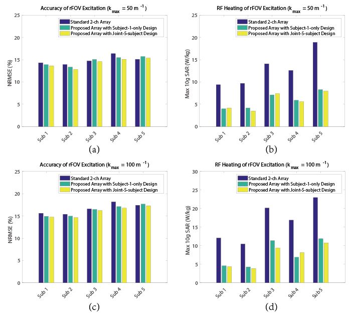
Figure 5 shows the excitation accuracy and maximum 10g SAR of different array design methods. The proposed 8-loop-2-channel array shows large reduction of 10g SAR as much as 50% than the standard 2-coil array. According to IEC regulations (11), the latter would exceed the maximum 10g SAR limit of 10W/kg, while the former would not. Again, the joint-5-subject design showed overall better performance than the subject-1-only design, signifies the importance of using a population of models. Coil combination weights and pruning strategy was also found to be robust on different excitation k-space trajectories.
Conclusion
Here a novel pTx array design method is proposed that integrates pTx RF pulse principles. It shows better excitation accuracy, lower SAR energy deposition, and robustness across different subjects and pulse parameters.Acknowledgements
This work was supported by NIH grant R01 EB016695.References
[1] Mooiweer, R., Sbrizzi, A., Raaijmakers, A. J.E., van den Berg, C. A.T., Luijten, P. R. and Hoogduin, H. (2016), Combining a reduced field of excitation with SENSE-based parallel imaging for maximum imaging efficiency. Magn. Reson. Med.. doi:10.1002/mrm.26346
[2] Lattanzi, R., Sodickson, D. K., Grant, A. K. and Zhu, Y. (2009), Electrodynamic constraints on homogeneity and radiofrequency power deposition in multiple coil excitations. Magn. Reson. Med., 61: 315–334. doi:10.1002/mrm.21782
[3] Cao, Z., Yan, X. and Grissom, W. A. (2016), Array-compressed parallel transmit pulse design. Magn. Reson. Med., 76: 1158–1169. doi:10.1002/mrm.26020
[4] Cao, Z., Yan, X. and Grissom, W. A. Proceedings of ISMRM 2016 in Singapore (Poster 2155).
[5] Christ A, Kainz W, Hahn EG, Honnegger K, Zefferer M, Neufeld E, Rascher W, Janka R, Bautz W, Chen J, Kiefer B, Schmitt P, Hollenbach HP, Shen J, Oberle M, Szczerba D, Kam A, Guag JW, Kustler N. The virtual family development of surface-based anatomical models of two adults and two children for dosimetric simulations. Phys Med Biol 2010; 55: N23–N38.
[6] Eichfelder, G. and Gebhardt, M. (2011), Local specific absorption rate control for parallel transmission by virtual observation points. Magn. Reson. Med., 66: 1468–1476. doi:10.1002/mrm.22927.
[7] Marques, J.P. and Bowtell, R. (2005), Application of a Fourier-based method for rapid calculation of field inhomogeneity due to spatial variation of magnetic susceptibility. Concepts Magn. Reson., 25B: 65–78. doi:10.1002/cmr.b.20034
[8] Malik, S. J., Keihaninejad, S., Hammers, A. and Hajnal, J. V. (2012), Tailored excitation in 3D with spiral nonselective (SPINS) RF pulses. Magn. Reson. Med., 67: 1303–1315. doi:10.1002/mrm.23118
[9] Cloos MA, Boulant N, Luong M, Ferrand G, Giacomini E, Hang MF, Wiggins CJ, Le Bihan D, Amadon A. Parallel-transmission-enabled magnetization-prepared rapid gradient-echo T1-weighted imaging of the human brain at 7 T. Neuroimage 2012;62:2140–2150.
[10] Carluccio G, Erricolo D, Oh S, Collins CM. An approach to rapid calculation of temperature change in tissue using spatial filters to approximate the effects of thermal conduction. IEEE Trans Biomed Eng 2013;60:1735–1741.
[11] European Committee for Electrotechnical Standardization. Particular requirements for the safety of magnetic resonance equipment for medical diagnosis (IEC 60601-2-33:2008). Technical report, European Committee for Electrotechnical Standardization, Brussels, Belgium; 2002.
Figures