4291
Improved traveling wave efficiency in 7T human MRI using wireless local loop and dipole arrays1Institute of Imaging Science, Vanderbilt University, Nashville, TN, United States, 2Department of Radiology and Radiological Sciences, Vanderbilt University, Nashville, TN, United States, 3Department of Radiology and Biomedical Imaging, University of California San Francisco, San Francisco, CA, United States, 4UCSF/UC Berkeley Joint Graduate Group in Bioengineering, San Francisco, CA, United States, 5Department of Biomedical Engineering, Vanderbilt University, Nashville, TN, United States
Synopsis
Traveling-wave MRI has robust matching performance and capability for large field-of-view (FOV) imaging. However, the efficiency of traveling-wave MRI is much lower than conventional methods, which limits its application. One way to improve the efficiency is to place local wireless resonators around the subject. The feasibility of this approach has been demonstrated in previous works using a single small loop. However, it is not clear whether other kinds of coils (such as electric dipoles) can be used as local elements, and it is not clear how much the improvements can be maintained in human imaging using an array design. By using wireless local loop coil and transverse dipole arrays, the transmit efficiency (B1+) of traveling-wave MRI can be improved by 3.4-fold in the brain and 2-fold in the knee. The coil types (loops or dipoles) should be carefully chosen for brain or knee imaging to maximize the improvement since they exhibit different types of coupling to the TE11 mode, and the enhancement depends on the local body configuration
Introduction
Traveling-wave MRI, which uses relatively small and simple RF transmit/receive antennas, has robust matching performance and capability for large field-of-view (FOV) imaging1. However, the efficiency of traveling-wave MRI is much lower than conventional methods2, which limits its application. One way to improve the efficiency is to introduce additional materials into the magnet bore, such as dielectric filings3,4 or metamaterials5. An alternative simple method is to place local wireless resonators around the subject. The feasibility of this approach has been demonstrated in previous works using a single small loop6,7, where traveling wave efficiency was improved by ~15-fold. However, it is not clear whether other kinds of coils (such as electric dipoles) can be used as local elements, and it is not clear how much the improvements can be maintained in human imaging using an array design.Methods
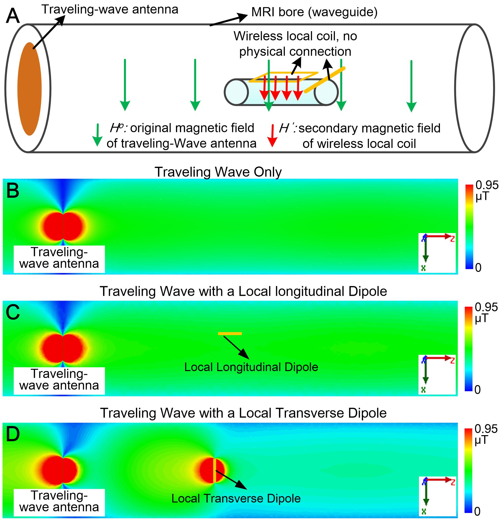
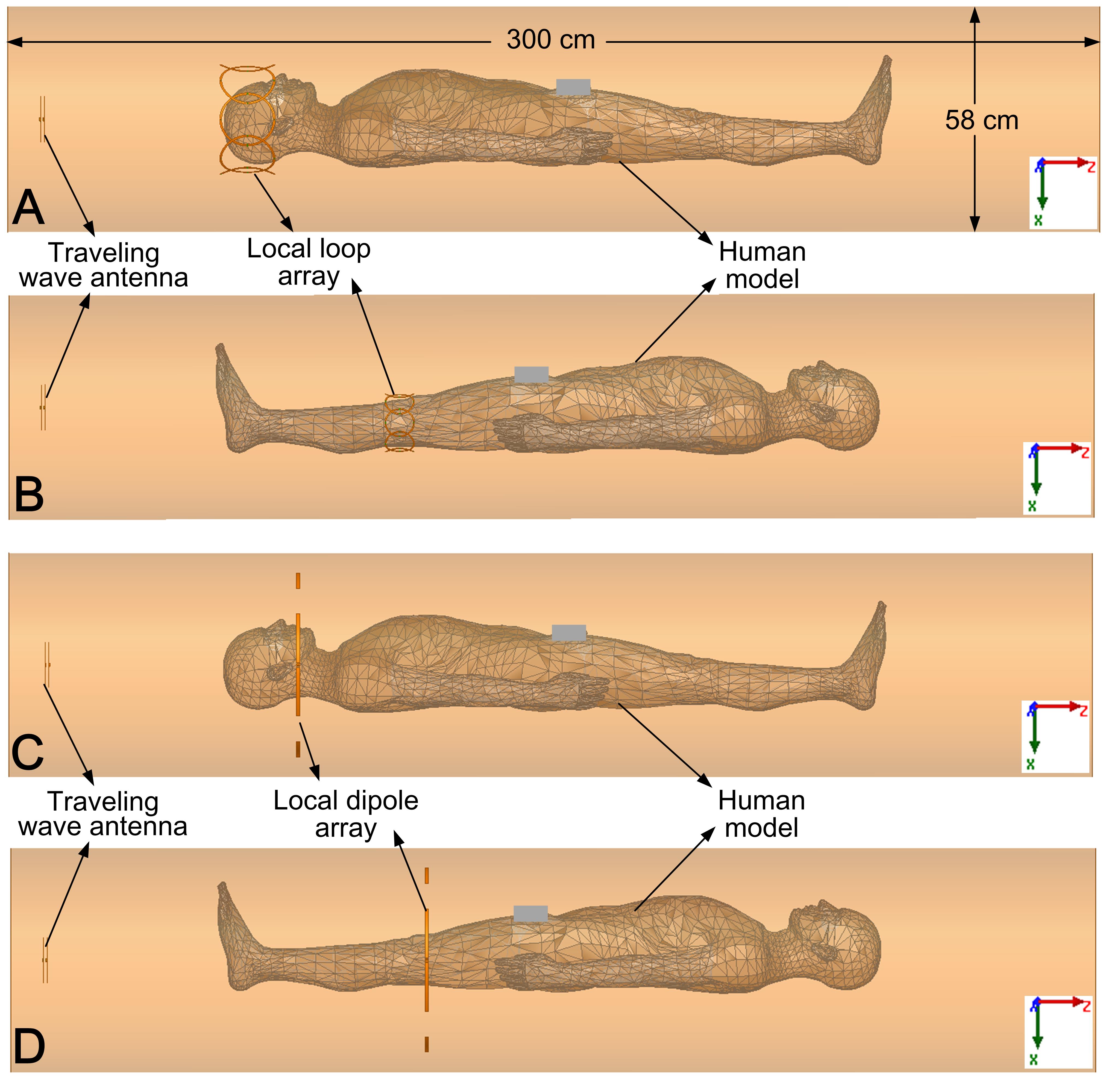
Fig. 1A illustrates a traveling wave MRI experiment with wireless local coils. The wireless coil produces a secondary magnetic field H’, which locally enhances B1 efficiency. To explore whether electric dipoles can locally enhance the B1 field, traveling-wave MRI was simulated in an empty bore with a local longitudinal (along the z-direction) dipole and a transverse dipole. Simulation results are shown in Figs. 1B-1D. It is found that the longitudinal dipole has no influence on the original B1+ field, but the transverse dipole can improve B1+ in front of it, which is expected since only the transverse dipole is coupled to the TE11 mode and acts as a reflector. Four local array setups were then examined in human body simulations: (I) brain with an 8-channel wireless loop array (Fig. 2A); (II) knee with an 8-channel wireless loop array (Fig. 2B); (III) brain with a 4-channel transverse dipole array (Fig. 2C); (Ⅳ) knee with a 4-channel transverse dipole array (Fig. 2D). Inductor-segmented crossed-dipoles were used as the traveling-wave antenna. Traditional traveling-wave setups without wireless coils were also simulated as comparisons.
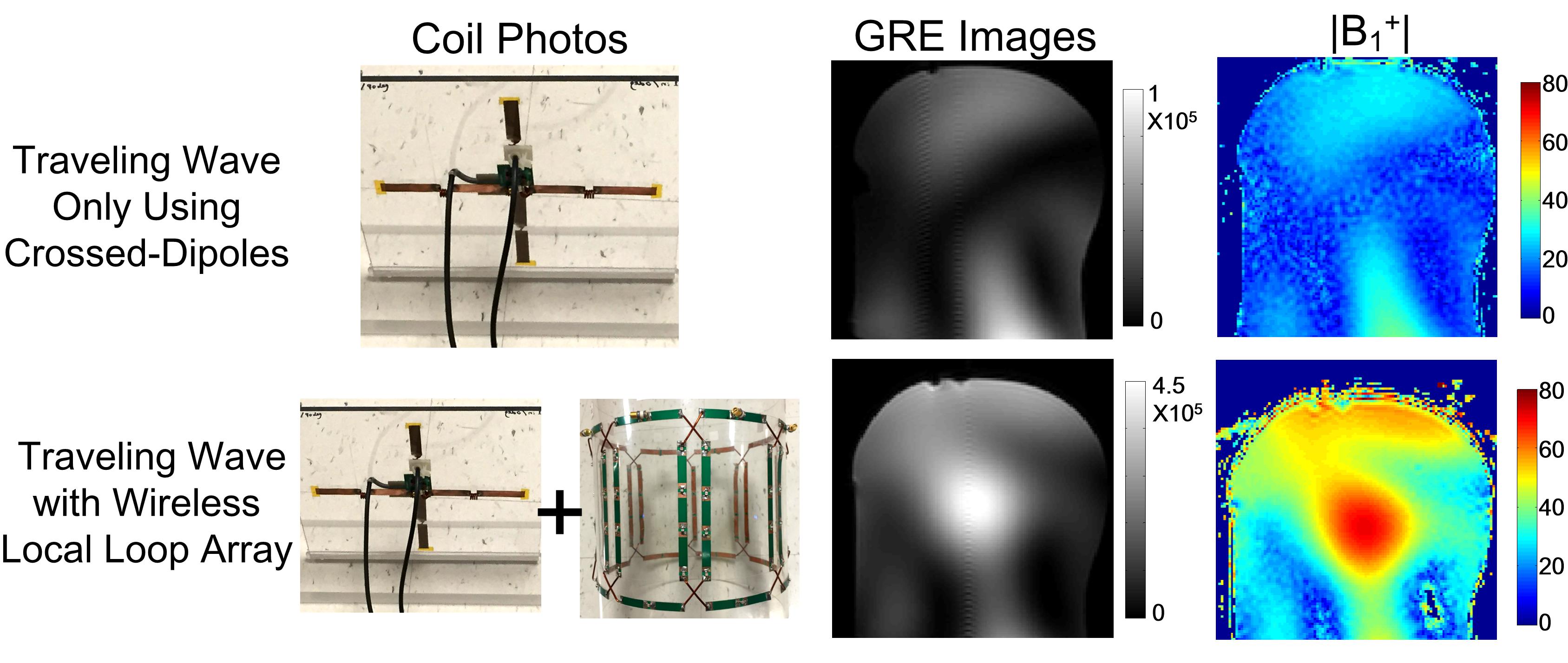
In all simulations, the total input power was set to 2 Watts. Simulations were performed using finite-element-method (Ansys HFSS, Canonsburg, PA, USA). To validate the simulation results, MR experiments were performed on a 7T whole-body scanner (Philips Healthcare, Best, Netherlands). GRE images and B1+ maps were acquired on a head-shaped phantom using the traveling-wave antenna with and without an 8-channel wireless local loop array. The parameters of the GRE sequence were: FOV=20x20 cm2, spatial resolution 2x2x5 mm3, TR/TE=1000/2.7 ms. B1+ maps were measured using DREAM method with the same input power8.
Results
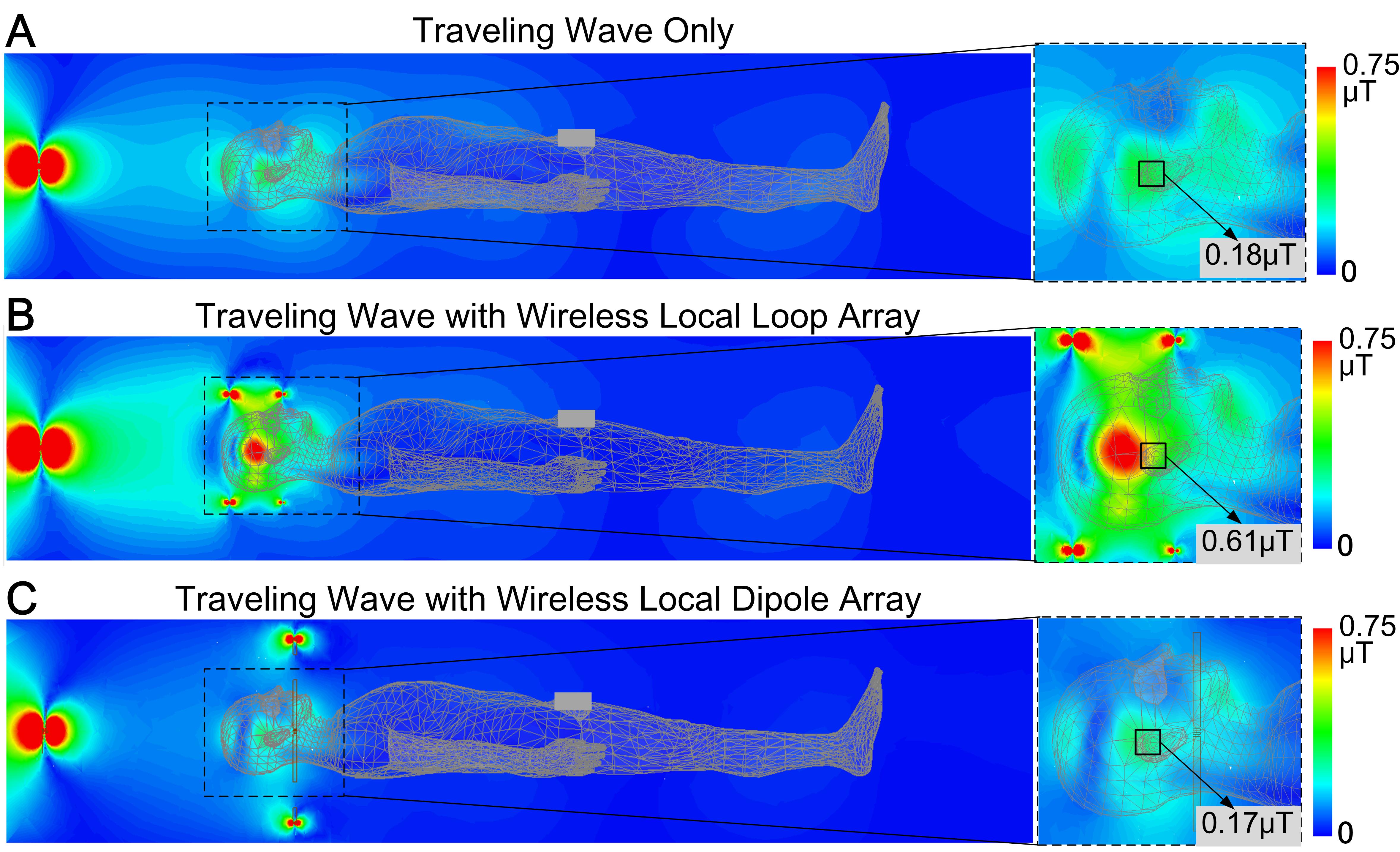
Fig. 3 shows the simulated B1+ efficiency in the brain. The wireless loop array increased the average B1+ field strength 3.4-fold. In the brain, most of the traveling wave transmit power is reflected by shoulders and wasted. With local loops, this reflection can be avoided since the most power deposits in the loops before arriving at the shoulders. For the wireless dipole array, however, the B1 efficiency is actually slightly lower than with traveling-wave alone, since the dipole array enhances this reflection effect.
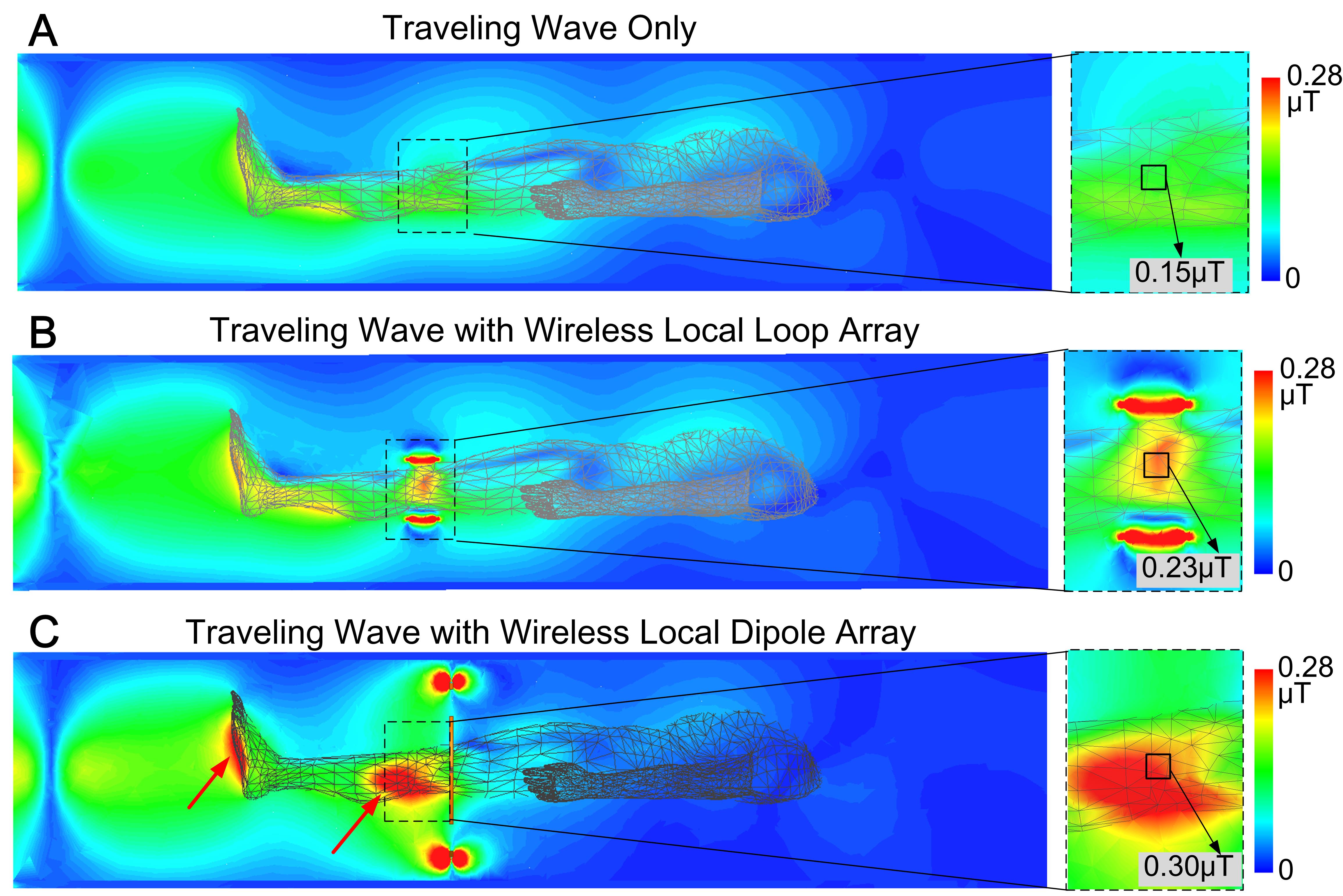
Fig. 4 shows the simulated B1+ efficiency in the knee. The local loop array resulted in a 1.5-fold increase in average B1+ field strength. Transmit power must travel across conductive tissues such as feet and legs before the knees. Therefore the improvement is relatively lower compared to the brain since much of the power is deposited before arriving at the knees. In comparison, the local dipole array increased the average B1+ field strength 2-fold, which is different from that in brain imaging. This is due to the large distance between the traveling wave antenna and the local dipole array, in which case the input RF signal and the reflected RF signal are deposited locally (red arrows in Fig. 4C).
Fig. 5 shows the traveling wave experimental results on a head-shaped phantom with and without an 8-channel loop array. As expected from the simulation results (Fig. 3B vs Fig. 3A), the B1+ efficiency increased approximately 3.5-fold using the local loop array.
Conclusion
By using wireless local loop coil and transverse dipole arrays, the transmit efficiency (B1+) of traveling-wave MRI can be improved by 3.4-fold in the brain and 2-fold in the knee. The coil types (loops or dipoles) should be carefully chosen for brain or knee imaging to maximize the improvement since they exhibit different types of coupling to the TE11 mode, and the enhancement depends on the local body configuration.Acknowledgements
This work was supported by NIH R01 EB 016695, NIH R21 EB 018521 and NIH R21 EB 020283.References
1. Brunner DO, Zanche ND, Frohlich J, Paska J, Pruessmann KP. Travelling-wave nuclear magnetic resonance. Nature. 2009;457:994–999
2. Zhang B, Sodickson DK, Lattanzi R, Duan Q, Stoeckel B, Wiggins GC. Whole body traveling wave magnetic resonance imaging at high field strength: Homogeneity, efficiency, and energy deposition as compared with traditional excitation mechanisms. Magn Reson Med. 67 (2012) 1183-1193 PMCID: PMC3376911
3. Andreychenko, A., Bluemink, J. J., Raaijmakers, A. J. E., Lagendijk, J. J. W., Luijten, P. R. and van den Berg, C. A. T. (2013), Improved RF performance of travelling wave MR with a high permittivity dielectric lining of the bore. Magn Reson Med, 70: 885–894. doi: 10.1002/mrm.24512.
4. Schmidt R and Webb, A improving travelling wave efficiency at 7 T using dielectric material placed ”beyond” the region of interest. Intl. Soc. Mag. Reson. Med. 2016; 3532
5. Zivkovic I., Scheffler K, Metamaterial cell for B1+ field manipulation at 9.4T MRI. Proc. Intl. Soc. Mag. Reson. Med. 2014; 4834
6. Zhang X, Pang Y, and Vigneron DB, SNR Enhancement by Free Local Resonators for Traveling Wave MRI, Proc. Intl. Soc. Mag. Reson. Med. 2014; 1357
7. Yan X, and Zhang X, Theoretical and simulation verification of SNR enhancement in traveling wave MRI using free local resonators, Proc. Intl. Soc. Mag. Reson. Med. 2016; 3535
8. Nehrke, K. and Börnert, P. (2012), DREAM—a novel approach for robust, ultrafast, multislice B1 mapping. Magn Reson Med, 68: 1517–1526. doi: 10.1002/mrm.24158
Figures