4288
Degree of RF MRI Coil Detuning and SAR Variations over an Anatomically Realistic Respiratory Cycle Modeled with the Finite Element Method1Worcester Polytechnic Institute, Worcester, MA, United States, 2NEVA Electromagnetics, LLC, Yarmouth Port, MA, United States
Synopsis
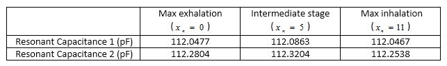
Respiratory motion is an important problem in Magnetic Resonance Imaging (MRI), contributing to image blurring during data acquisition and coil detuning. Using the concept of an ideal (perfectly matched and tuned at all available ports) RF transmit coil and the VHP-Female v4.0 dynamic CAD model, we estimate the detuning of a full-body RF coil detuning during the respiratory cycle. Our results show that the computed resonant capacitance values change by at most 0.5%.
Purpose
To estimate RF coil detuning during the normal respiratory cycle and determine ways to correct for respiratory artifactsMethod
An approximate method for modeling continuous respiratory motion in a CAD human virtual model subject to electromagnetic finite-element analysis has been realized. Its concept relies on using affine transformations (three-dimensional translations, rotations and scalings) to create polynomials of deformation for every structure involved in respiration, which are implemented in commercial FEM software packages in the form of a parametric sweep. This method does not require multiple copies of the CAD model or multiple project files. It enables use of arbitrary sampling times and an automatic reposition of on-body and in-body devices, such as medical implants. The method was applied to the Visible Human Project® (VHP)-Female phantom, a platform-independent, full-body triangular surface based electromagnetic computational model. Such an approach is not exact, but it may have sufficient accuracy when the parametric sweep is carefully designed. It will allow us to employ any temporal resolution, which is impossible with discrete models. To construct an anatomically relevant breathing cycle, we followed the anatomical data collected from Refs. [1-9] as close as possible.
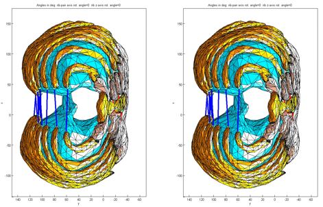
The user can initialize a discrete generic global variable,, define object geometry parameters as certain unique functions of , and then move/rotate/deform every object of a multi-object structure independently within the framework of the same project file, greatly facilitating large numeric studies that require these complex motions. In the present case, the 42 structures involved in respiration include 24 ribs, the heart, liver, lungs, stomach, skin shell, fat shell, 6 abdominal muscles, 2 erector spinae muscles, and 4 pectoralis muscles. No intersections between any structures are experienced as a result of these affine transformations. Figure 1 provides a depiction of minimum and maximum ribcage movement in the axial plane using this method. While air and total body volumes corresponding to these motions are automatically updated there is essentially no change in the mass of the virtual human model.
We next construct and simulate models mimicking maximum exhalation (), zero pressure gradient (), and maximum inhalation () using the perfectly matched RF coil model from Ref. [14]. This method uses a resonant-model of a coil optimally driven at all possible ports. The coil is loaded with a dynamic virtual human VHP-Female v. 4.0 Refs. [11, 12, 13].
Results
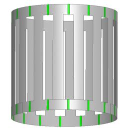
Table 1 shows averaged simulation results for the virtual human VHP-Female 4.0 in a high-pass fully body birdcage coil shown in Fig. 2 at the shoulder landmark. Our numerical results obtained with FEM software ANSYS Electronics Desktop reveal that detuning due to respiration is generally very small. The maximum deviation occurs in between maximum exhalation and maximum inhalation. A series of figures depicting Specific Absorption Rate (SAR) plots for various breathing stages is given in Table 2. Each tissue is augmented with accurate material properties.
Discussion and Conclusion
Finite-element simulations performed for the anatomically realistic breathing cycle provide quantitative estimates of RF coil detuning and SAR variations.Acknowledgements
No acknowledgement found.References
[1]. Siebenthal, M., “Analysis and Modelling of Respiratory Liver Motion using 4DMRI”, Ph.D. dissertation, Elect. Eng. and Inform. Technology Dept., ETH Zurich, Switzerland, 2008.
[2]. Grimm, R. et.al, “Self-gated MRI motion modeling for respiratory motion compensation in integrated PET/MRI,” Medical Image Analysis J., vol.19, pp., 110-120, 2015.
[3]. Lujan, A. et.al, “A method for incorporating organ motion due to breathing into 3D dose calculations,” Medical Physics, vol. 26, no. 5, pp. 715-720, 1999.
[4]. Lujan, A., Balter J., and Ten Haken R., “A method for incorporating organ motion due to breathing into 3D dose calculations in the liver: Sensitivity to variations in motion,” Medical Physics, vol.30, no.10, pp. 2643-2649, 2003.
[5]. Segars, W., Lalush D., and Tsui B., “Modelling respiration mechanics in the MCAT and spline-based MCAT phantom,” Nuclear Science Symp., Seattle, WA, 1999, vol. 2, pp. 985-989.
[6]. Wang, Y., Riederer S., and Ehman R., “Respiratory motion of the heart: Kinematics and the implications for the spatial resolution in coronary imaging,” Magnetic Resonance in Medicine, vol. 33, no. 5, pp.713-719, 1995.
[7]. West, J., Respiratory Physiology, 5th ed. Baltimore, Williams and Wilkins, 1995.
[8]. Zeng, R., “Estimating Respiratory Motion from CT Images via Deformable Models and Priors,” Ph.D. dissertation, Elect. Eng. Dept., University of Michigan, Ann Arbor, Michigan, 2007.
[9.] Eom, J. et.al, “Predictive modeling of lung motion over the entire respiratory cycle using measured pressure-volume data, 4DCT images, and finite element analysis,” Medical Physics, vol. 37, no.8, pp. 4389 – 4400, 2010.
[10]. Lemdiasov, R, Obi A, Ludwig R. A Numerical Postprocessing Procedure for Analyzing Radio Frequency MRI Coils. Concepts in Magnetic Resonance Part A. 2011; 38A(4): 133–147.
[11] Tran, A., Noetscher G., Louie S., Prokop A., Nazarian A., and Makarov S., “FEM Human Body Model with Embedded Respiratory Cycles for Antenna and E&M Simulations,” 2016 Antenna Application Symposium, Allerton Park, Monticello, IL, Sep. 20-22 2016
[12]. Yanamadala, J., Noetscher G., Louie S., Prokop A., Kozlov M., Nazarian A., and Makarov S, “Multi-Purpose VHP-Female Version 3.0 Cross-Platform Computational Human Model,” 10th European Conference on Antennas and Propagation 2016 (EuCAP16), Davos, Switzerland, April 10-15 2016.
[13]. Tankaria, H., Jackson X., Borwankar R., Srichandhru G., Tran A., Yanamadala J., Noetscher G., Nazarian A., Louie S., and Makarov S., “VHP-Female Full-Body Human CAD Model for Cross-Platform FEM Simulations – Recent Development and Validations,” 38th Annual Int. Conf. of the IEEE Engineering in Medicine and Biology Society (EMBC 2016), Orlando, FL, Aug. 16-20 2016.
[14]. Makarov, S., Tankaria H., Bogdanov G., Louie S., Burnham E., Noetscher G., “Generic Model of a Perfectly Matched RF Coil Intended for Regulatory SAR Computations with Virtual Human Models,” ISMRM 2017, Honolulu, Hi, April 22-27 2017
Figures