4277
Evaluation of aging effects on cerebral hemodynamics by Magnetic Resonance Imaging1Physics, University of Sao Paulo, Ribeirao Preto, Brazil
Synopsis
Recently, the effects of aging on human brain tissue, mainly how structural changes may be related to functional changes, have been extensively discussed. However, there is still no agreement on which brain regions have altered perfusion and how it is related with dementia. Therefore, in the present study, we investigated regional changes in perfusion and gray matter concentration in healthy aging, Mild Cognitive Impairment and Alzheimer’s disease. The results indicate a significant age- and disease-related reduction of regional cerebral perfusion associated with brain atrophy. Therefore, these alterations may be important biomarkers for neurodegeneration.
Purpose
During its development, the brain undergoes a series of anatomical, functional and organizational changes necessary to support the complex adaptive behavior of a normal individual fully mature. An approach to study these changes is the measurement of regional glucose metabolic use in different ages, or a physical quantity correlated with brain metabolism and local functional activity, such as the cerebral perfusion, which can be quantified by measuring the blood volume flowing through the vascular network in a particular region1. Therefore, the purpose of the present study is to investigate regional perfusion and brain atrophy, using Magnetic Resonance Imaging (MRI) techniques, in four subject groups: healthy young adults, healthy elderly adults, patients with Mild Cognitive Impairment (MCI) and patients with Alzheimer’s disease (AD).Methods
Twenty-four subjects were included in this study: six young healthy controls (3M/3F, mean age = 25.3±3.5 years), six elderly healthy controls (3M/3F, mean age = 68.8 ±2.9 years), six MCI patients (2M/4F, mean age = 72.2±8.6 years) and six early stage AD patients (1M/5F, mean age = 72.3±9.2 years). MRI were acquired in a Philips Achieva 3T System (Philips Achieva, The Netherlands), using a head coil for signal reception. For perfusion evaluation, axial images of Pulsed Arterial Spin Labeling (PASL) were acquired with the following parameters: 2D EPI, TR/TE = 4000/20 ms, flip angle = 90°, FOV = 240x240 mm², matrix = 80x80, 12 5-mm slices, post-labeling delay = 1500 ms, 40 dynamic pairs. For anatomical reference and Gray Matter (GM) atrophy analysis, images were acquired using a 3D T1-weighted gradient-echo sequence, with the following parameters: TR/TE = 7/3 ms, flip angle = 8°, matrix = 240 x 240, FOV = 240 x 240 mm², 160 1-mm slices. Images were processed using own routines developed in MATLAB (MathWorks, Natick, MA) and SPM12 routines. For PASL images, motion correction, spatial smoothing, coregistration to anatomical images and normalization to MNI space were applied. Perfusion-weighted images were obtained by subtracting each pair (control/tag) of PASL images. GM concentration was estimated using Voxel Based Morphometry (VBM) analysis on 3DT1 images. To compare groups, student’s t-tests were performed with FDR correction and significance set to p < 0.05.Results
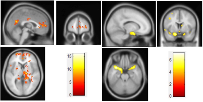
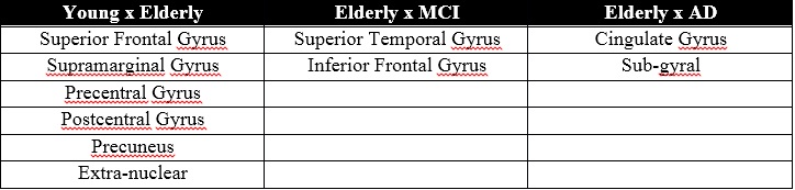
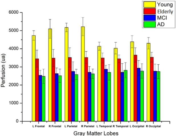
Voxel-wise analysis comparing young and elderly healthy adults showed age-related reduction in perfusion and GM concentration in different brain regions (Figure 1). Moreover, MCI and AD patients also presented reduced perfusion and GM concentration when compared to age-matched controls (Figure 2). Figure 3 shows brain regions with alterations in both parameters. A clusters-wise analysis comparing brain lobes was also performed, and indicated age- and disease-related reduction in perfusion mainly in frontal and parietal lobes (Figure 4).Discussion
Much has been discussed in recent years in relation to the effects of aging on human brain tissue mainly how structural changes may be related to functional changes. However, there is still no agreement in relation to which brain regions have altered perfusion and how it is related with diseases that are very common in elderly, such as MCI and AD. Therefore, in the present study, we investigated regional changes in perfusion and GM concentration in healthy aging and dementia. Comparing to young adults, healthy elderly showed reduced perfusion in different brain regions, in agreement with previous studies using ASL methods3. Reduced GM concentration was also observed. These alterations may be related to cognitive changes, mainly related to language and memory, reported for elderly adults, even in the absence of dementia4,5,6. So, some of these regions may be associated with vulnerability to neurological disorders in elderly. It is reinforced by the results with MCI and AD patients, which presented the same alterations, mainly in frontal regions. Therefore, perfusion alterations and GM atrophy may be important biomarkers for neurodegeneration. However, more data is being acquired to better investigate the relationship between these alterations and age-related cognitive decline.Conclusion
So far, the results of the present study indicate a significant reduction of cerebral perfusion associated with brain atrophy in some brain regions, in healthy aging and also in pre-dementia (MCI) and neurodegenerative disease (AD). However, not all regions with flow reduction presented regional atrophy. Further analysis and greater group sizes with allow more investigation on the relationship between brain perfusion and atrophy with healthy aging and early stages of dementia.Acknowledgements
No acknowledgement found.References
1. Liu Y, Zhu X, Feinberg D, et al. Arterial spin labeling MRI study of age and gender effects on brain perfusion hemodynamics. Magn Reson Med 2012;68:912-922.
2. Montagne A, Nation DA, Pa J, et al. Brain imaging of neurovascular dysfunction in Alzheimer’s disease. Acta Neuropathol 2016;131:687.
3. Liu Y, Zhu X, Feinberg D, et al. Arterial spin labeling MRI study of age and gender effects on brain perfusion hemodynamics. Magn Reson Med 2012;68(3):912-922.
4. Popa-Wagner A, Buga AM, Popescu B, Muresanu D. Vascular cognitive impairment, dementia, aging and energy demand. A vicious cycle. J Neural Transm 2013.
5. Ferreira LK, Regina ACB, Kovacevic N, et al. Aging effects on whole-brain functional connectivity in adults free of cognitive and psychiatric disorders. Cerebral Cortex 2015;1-15.
6. Peters R. Ageing and the brain. Postgrad Med J 2006;82(964):84–88.
Figures