4259
Oscillating Gradient Spin Echo Diffusion Tensor MRI of the Corpus Callosum with Typical Aging1Biomedical Engineering, University of Alberta, Edmonton, AB, Canada
Synopsis
This study compared short diffusion time (4 ms) oscillating gradient spin echo (OGSE) relative to regular long diffusion time (40 ms) pulsed gradient spin echo (PGSE) DTI in healthy individuals over a 40 year age span (n=12, 24 to 67 years old) to identify unique microstructure organization and reveal potential age related changes in three portions of the corpus callosum. Diffusivity and anisotropy were shown to depend on the diffusion time, mostly in the splenium, and these changed with age possibly reflecting alterations of restrictive dimensions such as the loss of specific axon diameters with aging.
PURPOSE
Oscillating gradient spin echo (OGSE) waveforms enable much shorter diffusion times (Δeff = 4 ms) than the typical pulsed gradient spin echo (PGSE, Δeff = 40 ms). Thus OGSE is sensitive to water diffusion restriction/hindrance over smaller length scales. We have previously shown that diffusion parameters derived from OGSE differ relative to PGSE in the white matter of young adults1. Large population studies have shown increases of mean diffusivity (MD) and decreases of fractional anisotropy (FA) with aging in adulthood2, but this has been measured with the usual long diffusion times. The purpose here was to evaluate whether short diffusion time OGSE DTI could capture unique age-related variability in sub-regions of the corpus callosum of healthy individuals.METHODS
OGSE 50 Hz (Δeff = 4 ms) and PGSE (Δeff = 40 ms) DTI was acquired in 12 healthy volunteers aged 24-67 years (7 males) on a Varian Inova 4.7T MRI using: scan time of either 5 min each for 20 axial slices over 5 cm centered on the corpus callosum (n=7) or 10 min each for 40 slices over 10 cm (n=5); 2D single-shot EPI with R=2 GRAPPA (4 element receive coils); TR=12.5 s; TE=110 ms; FOV=24 cm; 2x2x2.5 mm3; 6 averages; b=300 s/mm2; 6 gradient directions. The diffusion tensor data was processed using ExploreDTI. The OGSE FA map was non-linearly registered to the FMRIB58 1mm FA atlas using FSL, which was then applied to OGSE and PGSE FA, MD, axial diffusivity (AD) and radial diffusivity (RD) maps. AD, RD, MD, and FA were measured in the genu, body and splenium of corpus callosum using ROIs from the JHU atlas. All FA and diffusion parameters obtained with either OGSE or PGSE were first compared using a paired t-test in the three sub-regions of the corpus callosum. Differences between OGSE and PGSE were evaluated across the three regions with a one-way ANOVA with Tukey post-hoc test. The raw values for OGSE and PGSE were also assessed for linear correlations versus age (p<0.05).RESULTS
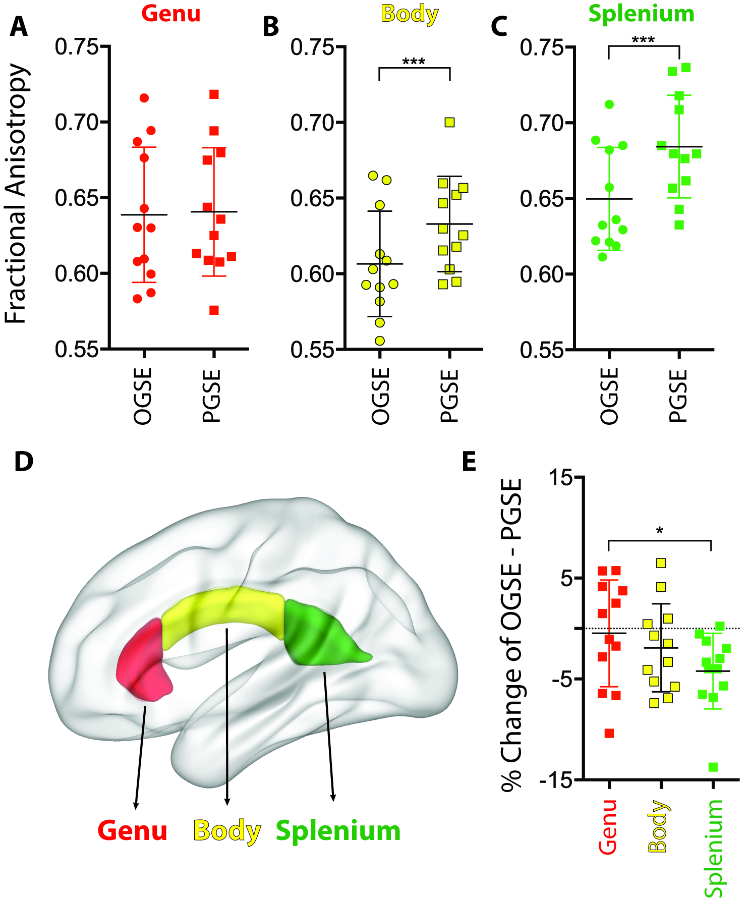
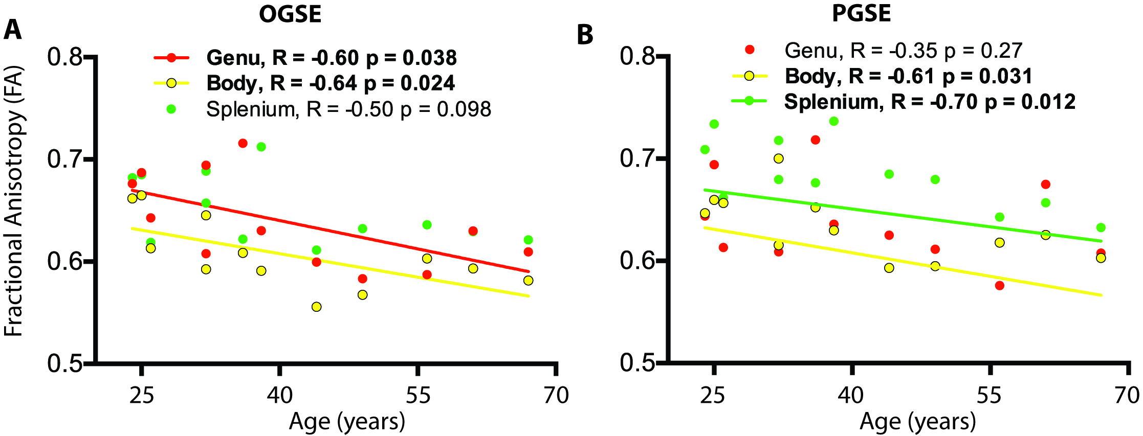
Diffusivity parameters (MD, RD, AD) were significantly higher with the short diffusion time OGSE when compared to the long diffusion time PGSE for all corpus callosum subregions ranging from 4% to 16% with the highest increase seen in the splenium. FA was lower for OGSE compared to PGSE in the body (0.607 ± 0.035 vs 0.633 ± 0.032, p<0.001) and in the splenium (0.650 ± 0.034 vs 0.684 ± 0.034, p<0.001) (Figure 1 for FA). Over a 40 year-span, FA decreased with age in genu (OGSE, R=-0.60, p=0.038), body (OGSE, R=-.0.64, p=0.024 and PGSE, R=-.0.61, p=0.031), and splenium (PGSE, R=-0.70, p=0.012), RD increased with age in genu (OGSE, R=0.60, p=0.040), body (OGSE, R=0.57, p=0.051, and PGSE, R=0.65, p=0.024), and splenium (PGSE, R=0.68, p=0.014) and MD increased with age in splenium (PGSE, R=0.61, p=0.035) (Figure 2 for FA).DISCUSSION AND CONCLUSIONS
The comparison of short diffusion time OGSE DTI to long diffusion time PGSE DTI reveals corpus callosum sub-regional variability showing that the splenium has the most robust OGSE/PGSE differences, thus suggesting unique micro-structural composition. These observations seem to support histological studies3,4 and MRI5 study that previously have shown discontinuity in the axonal size across the corpus callosum, where the splenium appears to have a higher proportion of larger axons. Regular long diffusion time DTI has in the past yielded greater changes in routine DTI parameters (e.g. FA) with age in frontal white matter regions such as the genu2. The fact that OGSE increases in radial diffusivity to a greater extent with age than PGSE in the genu indicates that water molecules are experiencing less barriers at the short diffusion time, more so in the older subjects. This could reflect the dominance of larger restrictive spaces at older age due to perhaps the loss of smaller axons. OGSE DTI using shorter diffusion times than other approaches appears to provide new insight into brain changes with aging and could validate histological findings of age/fiber size associations in corpus callosum sub-regions6 as well as providing a new tool for clinical applications.Acknowledgements
Canadian Institutes of Health Research (CIHR)References
1. Baron CA, Beaulieu C. Oscillating gradient spin-echo (OGSE) diffusion tensor imaging of the human brain. Magn Reson Med 2014; 72:726-736.
2. Lebel C, Caverhill-Godkewitsch S, Beaulieu C. Age-related regional variations of the corpus callosum identified by diffusion tensor tractography. Neuroimage. 2010 Aug 1;52(1):20-31.
3. Aboitiz F, Scheibel AB, Fisher RS, Zaidel E. Fiber composition of the human corpus callosum. Brain Res. 1992 Dec 11;598(1-2):143-53.
4 Lamantia AS, Rakic P. Cytological and quantitative characteristics of four cerebral commissures in the rhesus monkey. J Comp Neurol. 1990 Jan 22;291(4):520-37.
5. Zhang H, Hubbard PL, Parker GJ, Alexander DC. Axon diameter mapping in the presence of orientation dispersion with diffusion MRI. Neuroimage. 2011 Jun 1;56(3):1301-15
6. Aboitiz F, Rodríguez E, Olivares R, Zaidel E. Age-related changes in fibre composition of the human corpus callosum: sex differences. Neuroreport. 1996 Jul 29;7(11):1761-4.
Figures