4239
Minimizing the confounding effect of gadolinium contrast on subsequent ferumoxytol MRI in the brain1Dept. of Radiology, Oregon Health and Science University, Portland, OR, United States, 2Oregon Health and Science University, Portland, OR, United States, 3Dept. of Neurology, Oregon Health and Science University, Portland, OR, United States
Synopsis
Ferumoxytol as an MRI contrast agent has the advantage of long intravascular phase and strong transverse relaxivity, allowing high resolution depiction of abnormal vasculature and steady state blood volume (SS-CBV) mapping. In clinical studies it would be beneficial to use ferumoxytol as an immediate extension of standard of care gadolinium enhanced MRI, instead of imaging on two separate days. This study concludes that the 3D T2* weighted acquisition has substantial T1 weighting, while the 2D acquisition does not, therefore the latter one is preferable for ferumoxytol vascular imaging if gadolinium is on board.
Background and Purpose
Gadolinium enhanced MRI is the standard of care for various CNS pathologies. Ferumoxytol, used off label as an MRI contrast agent can visualize abnormal vascularity and can be used to create high resolution steady state CBV maps, which are not feasible with standard gadolinium.1, 2 Clinically, it would be beneficial to extend gadolinium MRI with subsequent ferumoxytol imaging in the same MRI session, with no confounding effects between the two contrast agents.
The purpose of this study was to test how gadolinium administration alters the signal on various T2*-weighted images, which may be used for steady state CBV mapping with ferumoxytol.
Methods and Materials
Fourteen cases of 9 subjects with suspected glioblastoma, enrolled in an ongoing imaging trial (eIRB# 9846) were analyzed. In each case T2* weighted images were acquired on two consecutive days: On day1, pre and post 0.1mmol/kg standard dose of gadoteridol, on day2 pre and post 7mg/kg (up to 510mg total) ferumoxytol. Subjects were scanned using a 3T Philips scanner. Two types of sequences, were used: the magnitude images of modified susceptibility weighted imaging (SWI, 3D acquisition, TR/TE/FA: 26/20/15, FOV:210x200mm2 acq matrix 300x300, 2mm thickness, overlap 1mm, scan time: 214s) and multi echo fast field echo (mFFE, 2D acquisition, TR 920ms, five echo times between 6.9 and 29 ms, FA:18, FOV 230x184mm2, acq matrix 384x307, 4mm thickness, 1mm gap, scan time: 227s.) Images from the 5 echoes were analyzed separately as well as averaged. Signal intensity changes within the enhancing tumor, (normalized to the cerebrospinal fluid in the lateral ventricles) were evaluated between pre and post gadolinium, as well as pre and post ferumoxytol. Ratios of post/pre contrast values were analyzed using one sample T-tests of the SWI data as well as the mFFE averaged images. Additionally the signal changes at various echo times were also described.Results
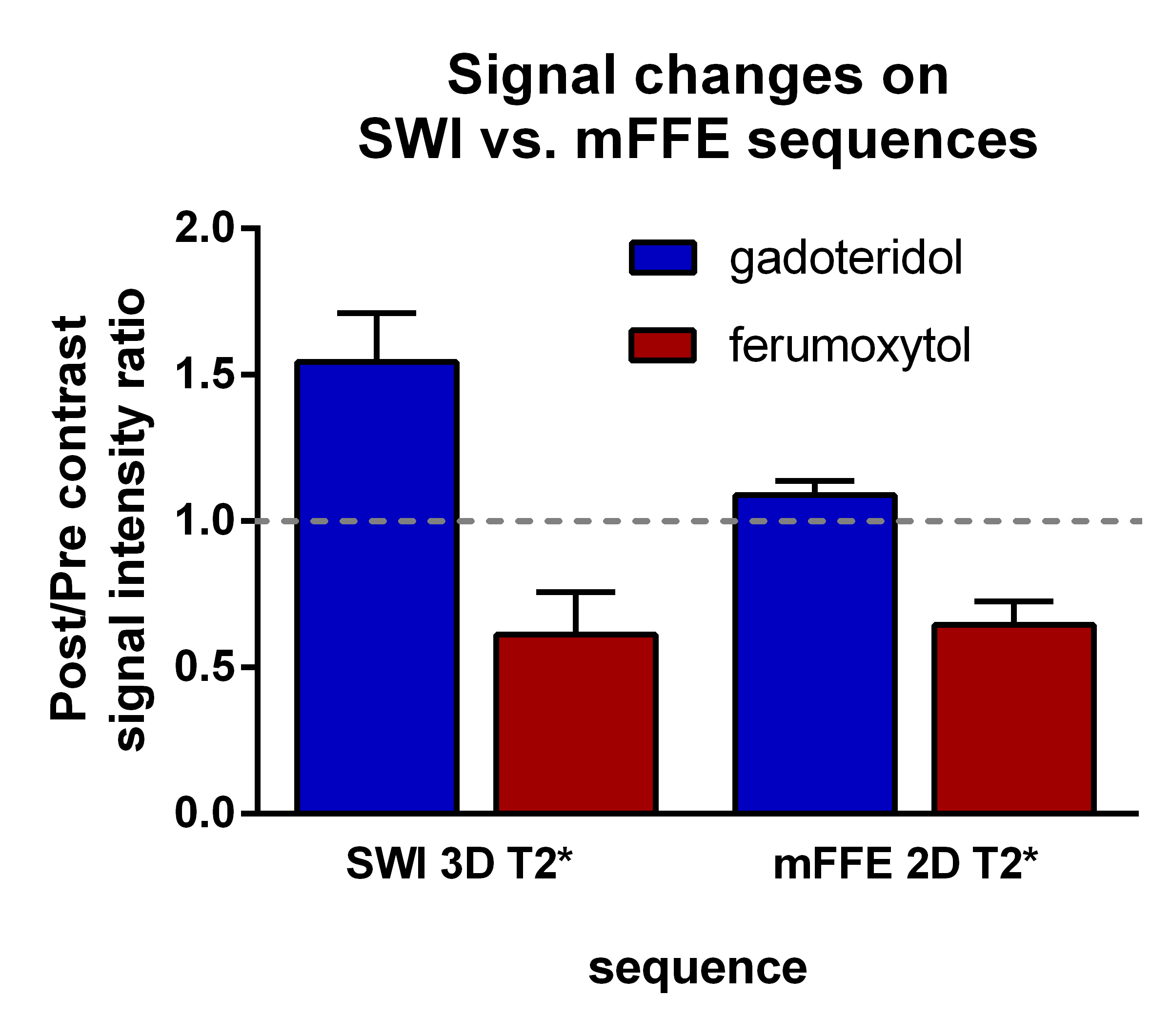
Signal intensity ratios post/pre gadolinium increased on SWI 3D T2*-weighted scans substantially to 1.54±0.29 (Mean ± SD) p < 0.0001, while only minimally on the mFFE 2D T2*-weighted scans to 1.09±0.08, p=0.0014. (figure 1.) Ferumoxytol caused signal drop on both sequences similarly, to 0.61±0.25 on the SWI 3D and 0.65±0.14 on the mFFE 2D acquisitions (p < 0.0001). Figure 2 shows the post/pre contrast ratios at various echo times using the mFFE dataset. Increasing echo times only caused decreasing signal ratio with ferumoxytol, but remained constant with gadoteridol.Discussion
Optimizing acquisition for SS-CBV mapping or visualization of abnormal vasculature using ferumoxytol is especially important when two contrast agents are administered in the same imaging session. Minimizing the confounding effect from prior administered gadolinium based agents by using sequences with minimal T1 sensitivity can eliminate errors from constantly changing gadolinium enhancement. Although in general 3D acquisition of T2* weighted imaging (as used in SWI) has the benefit of thin slices and therefore superior multi-planar reconstruction, its T1 sensitivity is substantial. 2D acquisition is preferable in studies performed if gadolinium is on board. Echo time had no effect on the signal ratio post/pre gadolinium, therefore echo time is unlikely a crucial parameter in minimizing T1 effects of gadolinium.Conclusion
Given the predominant T1 relaxation time shortening effect of gadolinium and T2* shortening effect of ferumoxytol early after injection, mFFE 2D T2*-weighted acquisition lacking substantial T1 effects is the best choice to image the intravascular space with ferumoxytol when added to standard of care gadolinium MRI. This finding should be considered when designing dual agent clinical studies or when using ferumoxytol off label subsequent to standard of care gadolinium MRI.Acknowledgements
No acknowledgement found.References
1. Varallyay, C.G., E. Nesbit, R. Fu, et al. High-resolution steady-state cerebral blood volume maps in patients with central nervous system neoplasms using ferumoxytol, a superparamagnetic iron oxide nanoparticle. J Cereb Blood Flow Metab, 2013. 33(5): p. 780-6.
2. Christen T, Ni W, Qiu D, et al. High-resolution cerebral blood volume imaging in humans using the blood pool contrast agent ferumoxytol. Magn Reson Med. 2013 Sep;70(3):705-10.
Figures
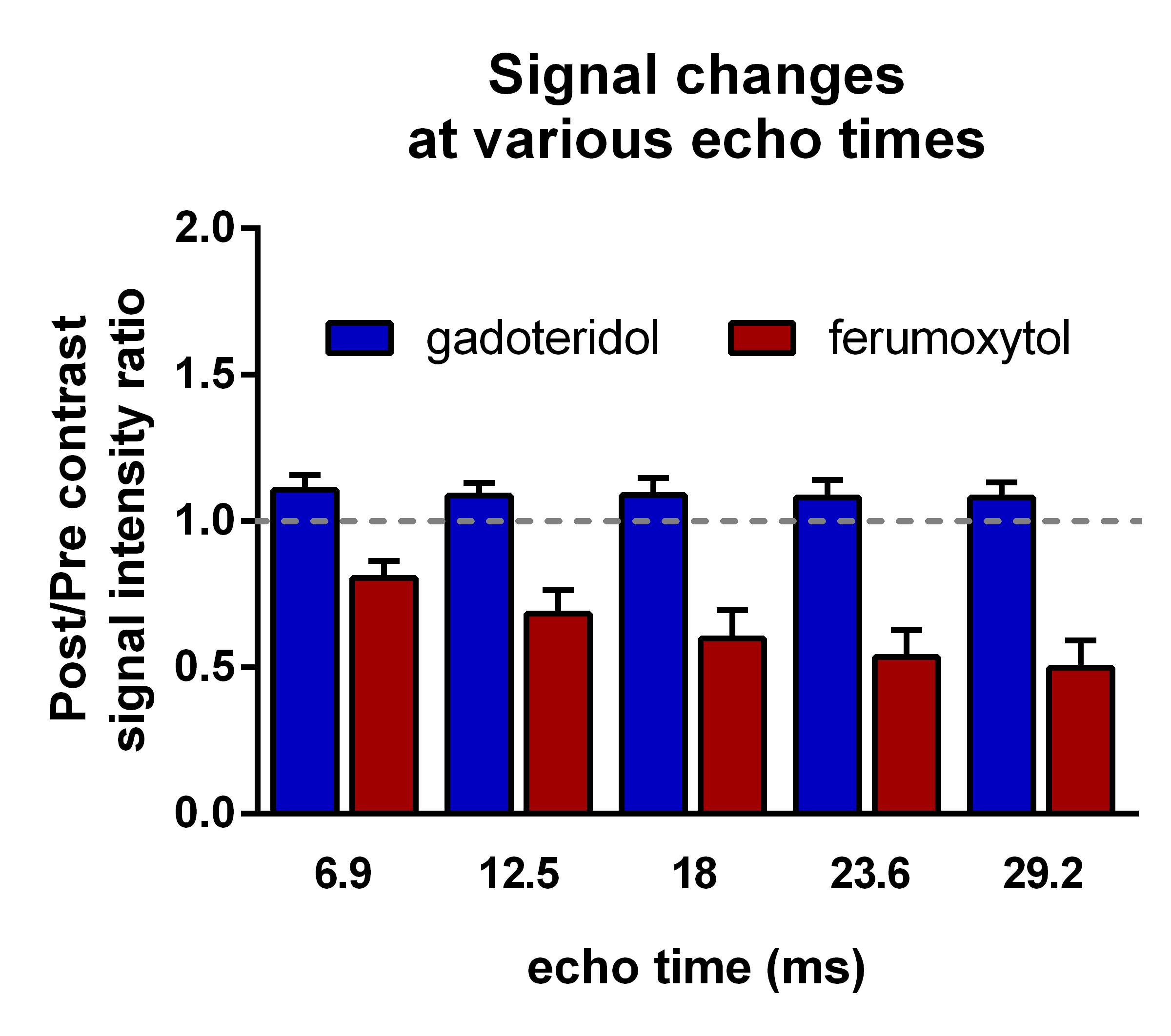
Means of post/pre contrast signal ratios in enhancing tumors at various echo times from the mFFE acquisition. n=14. Error bars indicate 95% CI. While increasing echo time result in decreasing signal ratios with ferumoxytol, there is no change in signal ratios after gadoteridol. This means that the T1 effect can not be compensated by altering the echo time.