4235
MRI Signal Enhancement in Early-Stage GBM Detection by Nonlinear Spin Dynamics using Active Feedback Fields1Chemistry and Biochemistry, University of California, Los Angeles, Los Angeles, CA, United States, 2Chemistry, National Taiwan University, Taipei, Taiwan
Synopsis
Application of MR imaging is very limited in imaging sensitivity and contrast in regards to the detection of brain tumor at an early stage because of tiny variations between healthy tissue and early-stage tumor. The radiation damping effect provides insight about the nonlinear spin evolution. Taking advantage of nonlinear spin evolution, we confirmed theoretically and experimentally that MR imaging provides a more stable and significant contrast in comparison to conventional methods.
Introduction
Studies have shown that the early-stage tumor shows a decrease of oxygen level in blood vessels leading to lower concentration of deoxyhemoglobin. This low concentration of deoxyhemoglobin causes the shift of local susceptibility between healthy tissue and brain tumor. Conventional longitudinal and transverse relaxation-based contrast mechanisms are relatively insensitive to these small variations, making it difficult to create significant contrast between healthy and tumor tissues. The radiation damping effect provides insight about the nonlinear spin evolution1. In addition to the deoxyhemoglobin-induced local magnetic field, an active feedback field was introduced to simulate the radiation damping effect. The transient magnetization is picked up and fed back to the active feedback field immediately, which generates avalanching spin amplification and enhances the weak dipolar-field perturbations from the deoxyhemoglobin. We confirmed theoretically and experimentally about sensitivity of this nonlinear spin evolution to subtle change of concentration of deoxyhemoglobin. This new MR imaging method produces a more stable and significant contrast in comparison to conventional methods. Thus, it enables more successful detections of medical diagnosis of early stage tumor.Method
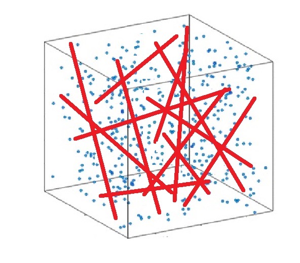
In-vivo experiment and Monte-Carlo simulation were carried out to test this method. In simulation, both healthy tissue and tumor tissue are modeled as voxels containing randomly orientated cylinders 2, 3 shown as Figure 1. More than 5000 water protons are randomly distributed inside the voxel and each of them performs the three dimensional random walk with fixed diffusion coefficient 4. The signal dephasing in each voxel dependents on the deoxyhemoglobin-induced local magnetic field variation, which in turn depends on the orientation of the vessel and the susceptibility difference5, 6. To model the contribution from bulk water protons that far away from the dipole center, more than 50 voxels without blood vessels are generated. To model the field inhomogeneity in human brain, different resonance offset associated with different voxels follows the Gaussian distribution. Experimental results were carried out using U87 glioblastoma tumor cell. Imaging were acquired on the 13th day after tumor cell injection.Results
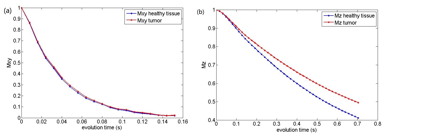
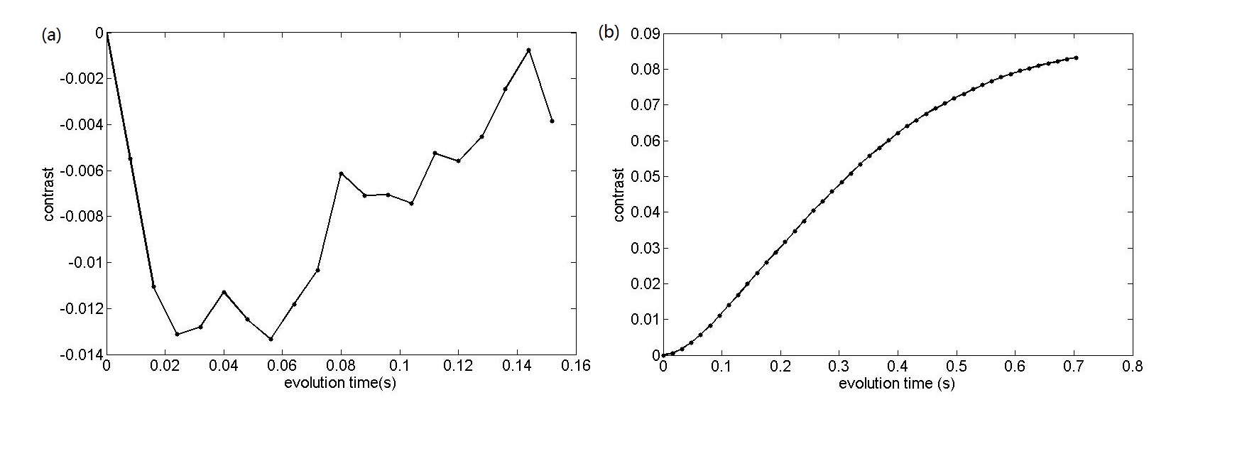
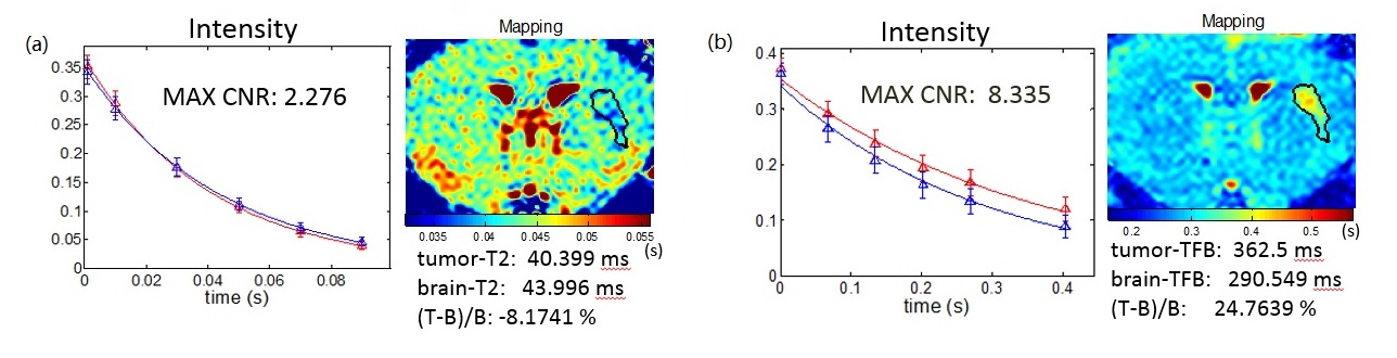
Figure 2 (a)(b) show the magnetization attenuation. Figure 3(a)(b) shows the simulation contrast. The contrast in conventional T2 weighted image vanishes rapidly. Moreover, the maximum value appear at 0.013, which usually is regarded as noise. A great enhancement in the contrast was obtained with the active field back field. The contrast accumulated through the nonlinear spin dynamics in present of active feedback and the maximum contrast appears at 0.095 which is more than 7.3 times higher. Figure 4(a)(b) shows the experiment results. The image acquired with active feedback field using homemade device showed a significant contrast while the images acquired from conventional T2 weighted imaging was insensitive to early stage brain tumor.Acknowledgements
This work was supported by the Camille and Henry Dreyfus Foundation (TC-05-053), National Science Foundation (DMS-0833863, CHE-1112574, and CHE-1416598), Hirshberg Foundation for Pancreatic Cancer Research, and Taiwan Ministry of Science and Technology (NSC 100-2113-M-002-008, NSC 101-2113-M-002-018, and MOST 103-2923-M-002-006).
References
1. Lin YY, Lisitza N, Ahn S, Warren WS. Resurrection of crushed magnetization and chaotic dynamics in solution NMR spectroscopy. Science 2000;290:118–121.
2. Hardy P, Henkelmen R M. On the transverse relaxation rate enhancement induced by Diffusion of Spins through inhomogeneous fields. Magn. Reson. Med. 1991; 17(2): 348-356.
3. Bouchet A, Bidart M, Miladi I, et al. Characterization of the 9L gliosarcoma implanted in the Fischer rat: an orthotopic model for a grade IV brain tumor. Tumor Biol. 2014; 35, 6221–6233.
4. Le Bihan, D. Looking into the functional architecture of the brain with diffusion MRI. Nat. Rev., Neurosci. 2003; 4(6): 469–480.
5. Springer C S, Xu Y, Aspects of bulk magnetic susceptibility in in vivo MRI and MRS. New developments in contrast agent research. Euro. Magn. Reson. Forum, Blonay, Switzerland. 1991; 13-25.
6. Pawlik, G, Rackl A, Bing RJ. Quantitative capillary topography and blood flow in the cerebral cortex of cats: an in vivo microscopic study. Brain Res. 1981; 208(1): 35-58.
Figures